Bolus tracking
Encyclopedia


Computed tomography
X-ray computed tomography or Computer tomography , is a medical imaging method employing tomography created by computer processing...
imaging, to visualise vessels more clearly. A bolus
Bolus (medicine)
In medicine, a bolus is the administration of a medication, drug or other compound that is given to raise its concentration in blood to an effective level...
of radio-opaque contrast media is injected
Injection (medicine)
An injection is an infusion method of putting fluid into the body, usually with a hollow needle and a syringe which is pierced through the skin to a sufficient depth for the material to be forced into the body...
into a patient via a peripheral intravenous cannula
Cannula
A cannula or canula is a tube that can be inserted into the body, often for the delivery or removal of fluid or for the gathering of data...
. Depending on the vessel being imaged, the volume of contrast is tracked using a region of interest at a certain level and then followed by the CT scanner
Computed tomography
X-ray computed tomography or Computer tomography , is a medical imaging method employing tomography created by computer processing...
once it reaches this level. Images are acquired at a rate as fast as the contrast moving through the blood vessels.
Applications
This method of imaging is used primarily to produce images of arteries, such as the aortaAorta
The aorta is the largest artery in the body, originating from the left ventricle of the heart and extending down to the abdomen, where it branches off into two smaller arteries...
, pulmonary
Pulmonary artery
The pulmonary arteries carry deoxygenated blood from the heart to the lungs. They are the only arteries that carry deoxygenated blood....
artery, cerebral and carotid
Carotid artery
Carotid artery can refer to:* Common carotid artery* External carotid artery* Internal carotid artery...
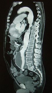
arteries. The image shown illustrates this technique on a sagittal MPR (multi planar reformat). The image is demonstrating the blood flow through an abdominal aortic aneurysm
Abdominal aortic aneurysm
Abdominal aortic aneurysm is a localized dilatation of the abdominal aorta exceeding the normal diameter by more than 50 percent, and is the most common form of aortic aneurysm...
or AAA. The bright white on the image is the contrast. You can see the lumen of the aorta in which the contrast is contained, surrounded by a grey 'sack', which is the aneurysm
Aneurysm
An aneurysm or aneurism is a localized, blood-filled balloon-like bulge in the wall of a blood vessel. Aneurysms can commonly occur in arteries at the base of the brain and an aortic aneurysm occurs in the main artery carrying blood from the left ventricle of the heart...
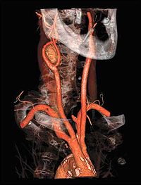
. Images acquired from a bolus track, can be manipulated into a MIP (maximum intensity projection
Maximum intensity projection
In scientific visualization, a maximum intensity projection is a volume rendering method for 3D data that projects in the visualization plane the voxels with maximum intensity that fall in the way of parallel rays traced from the viewpoint to the plane of projection...
) or a volume rendered image
Volume rendering
In scientific visualization and computer graphics, volume rendering is a set of techniques used to display a 2D projection of a 3D discretely sampled data set.A typical 3D data set is a group of 2D slice images acquired by aCT, MRI, or MicroCT scanner....
.
See also
- Computed tomography angiographyComputed tomography angiographyComputed tomography angiography is a computed tomography technique used to visualize arterial and venous vessels throughout the body. This ranges from arteries serving the brain to those bringing blood to the lungs, kidneys, arms and legs.-Technique:...
- CT pulmonary angiogramCT pulmonary angiogramCT pulmonary angiogram is a medical diagnostic test that employs computed tomography to obtain an image of the pulmonary arteries. Its main use is to diagnose pulmonary embolism...
- Endovascular surgeryEndovascular surgeryEndovascular surgery is a form of minimally invasive surgery that was designed to access many regions of the body via major blood vessels.Endovascular techniques were originally designed for diagnostic purposes. Basic techniques involve the introduction of a catheter percutaneously into a large...
- EVAR

