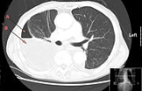
Empyema
Encyclopedia
Pleural empyema
(also known as a pyothorax or purulent pleuritis) is an accumulation of pus
in the pleural cavity
. Most pleural empyemas arise from an infection within the lung
(pneumonia
), often associated with parapneumonic effusions. There are three stages: exudative, fibrinopurulent and organizing. In the exudative stage, the pus accumulates. This is followed by the fibrinopurulent stage in which there is loculation of the pleural fluid (the creation of pus pockets). In the final organizing stage, scarring of the pleural space may lead to lung entrapment.
 Symptoms of pleural empyema may vary in severity. Typical symptoms include: cough
, fever
, chest pain
, sweating and shortness of breath
.
Clubbing may be present in cases of a chronic nature. There is a dull percussion note and reduced breath sounds on the affected side of the chest. Other diagnostic tools include a blood white cell count, chest x-ray
, CT scan, and ultrasonography
.
; frank pus or merely cloudy fluid may be aspirated from the pleural space. The pleural fluid typically has a leukocytosis, low pH (<7.20), low glucose (<60 mg/dL), a high LDH (lactate dehydrogenase), elevated protein and may contain infectious organisms.
may be inserted, often using ultrasound guidance. Intravenous antibiotic
s are given.
Chest tubes in the setting of pleural empyema have a tendency to be clogged by the thick pus. To combat this problem, surgeons will often place one or more large bore chest tubes. Insufficient drainage of the pleural empyema, particularly in loculated empyema, can lead to re accumulation of pus and infected material, a worsening clinical picture, organ failure and even death. Thus managing chest tube function is particularly important in the treatment of a pleural empyema. To improve the chest tube drainage, fibrinolytics and DNA enzyme can be given intrapleurally through the chest tube to break the fibrinous septation and to reduce the pus viscosity. Although these adjunct treatments are proven effective, its administration may cause rare but life-threatening intrapleural hemorrhage and hypersensitivity reaction.
If this is insufficient, surgical debridement
of the pleural space may be required. This is frequently done using video-assisted thoracoscopic techniques but if the disease is chronic, a limited thoracotomy may be necessary to fully drain the pus and remove the fibrinopurulent exudate from the lung and from the chest wall. Occasionally, a full thoracotomy, formal decortication and pleurectomy are required. Rarely, portions of the lung have to be resected.
An earlier form of treatment involved surgical removal of most of the ribs on the infected side of the thorax, causing a permanent collapse of the lung and obliteration of the infected pleural space. This would leave the patient with a large portion of the upper chest removed, giving the impression that the shoulder had been detached from the body. Rarely performed today, the surgery was common during World War I
.
Empyema
Pleural empyema is an accumulation of pus in the pleural cavity. Most pleural empyemas arise from an infection within the lung , often associated with parapneumonic effusions. There are three stages: exudative, fibrinopurulent and organizing. In the exudative stage, the pus accumulates...
(also known as a pyothorax or purulent pleuritis) is an accumulation of pus
Pus
Pus is a viscous exudate, typically whitish-yellow, yellow, or yellow-brown, formed at the site of inflammatory during infection. An accumulation of pus in an enclosed tissue space is known as an abscess, whereas a visible collection of pus within or beneath the epidermis is known as a pustule or...
in the pleural cavity
Pleural cavity
In human anatomy, the pleural cavity is the potential space between the two pleura of the lungs. The pleura is a serous membrane which folds back onto itself to form a two-layered, membrane structure. The thin space between the two pleural layers is known as the pleural cavity; it normally...
. Most pleural empyemas arise from an infection within the lung
Lung
The lung is the essential respiration organ in many air-breathing animals, including most tetrapods, a few fish and a few snails. In mammals and the more complex life forms, the two lungs are located near the backbone on either side of the heart...
(pneumonia
Pneumonia
Pneumonia is an inflammatory condition of the lung—especially affecting the microscopic air sacs —associated with fever, chest symptoms, and a lack of air space on a chest X-ray. Pneumonia is typically caused by an infection but there are a number of other causes...
), often associated with parapneumonic effusions. There are three stages: exudative, fibrinopurulent and organizing. In the exudative stage, the pus accumulates. This is followed by the fibrinopurulent stage in which there is loculation of the pleural fluid (the creation of pus pockets). In the final organizing stage, scarring of the pleural space may lead to lung entrapment.
Symptoms

Cough
A cough is a sudden and often repetitively occurring reflex which helps to clear the large breathing passages from secretions, irritants, foreign particles and microbes...
, fever
Fever
Fever is a common medical sign characterized by an elevation of temperature above the normal range of due to an increase in the body temperature regulatory set-point. This increase in set-point triggers increased muscle tone and shivering.As a person's temperature increases, there is, in...
, chest pain
Chest pain
Chest pain may be a symptom of a number of serious conditions and is generally considered a medical emergency. Even though it may be determined that the pain is non-cardiac in origin, this is often a diagnosis of exclusion made after ruling out more serious causes of the pain.-Differential...
, sweating and shortness of breath
Dyspnea
Dyspnea , shortness of breath , or air hunger, is the subjective symptom of breathlessness.It is a normal symptom of heavy exertion but becomes pathological if it occurs in unexpected situations...
.
Clubbing may be present in cases of a chronic nature. There is a dull percussion note and reduced breath sounds on the affected side of the chest. Other diagnostic tools include a blood white cell count, chest x-ray
X-ray
X-radiation is a form of electromagnetic radiation. X-rays have a wavelength in the range of 0.01 to 10 nanometers, corresponding to frequencies in the range 30 petahertz to 30 exahertz and energies in the range 120 eV to 120 keV. They are shorter in wavelength than UV rays and longer than gamma...
, CT scan, and ultrasonography
Medical ultrasonography
Diagnostic sonography is an ultrasound-based diagnostic imaging technique used for visualizing subcutaneous body structures including tendons, muscles, joints, vessels and internal organs for possible pathology or lesions...
.
Diagnosis
Diagnosis is confirmed by thoracentesisThoracentesis
Thoracentesis , also known as thoracocentesis or pleural tap, is an invasive procedure to remove fluid or air from the pleural space for diagnostic or therapeutic purposes. A cannula, or hollow needle, is carefully introduced into the thorax, generally after administration of local anesthesia...
; frank pus or merely cloudy fluid may be aspirated from the pleural space. The pleural fluid typically has a leukocytosis, low pH (<7.20), low glucose (<60 mg/dL), a high LDH (lactate dehydrogenase), elevated protein and may contain infectious organisms.
Treatment
Definitive treatment for pleural empyema entails drainage of the infected pleural fluid or pus. A chest tubeChest tube
A chest tube is a flexible plastic tube that is inserted through the side of the chest into the pleural space. It is used to remove air or fluid , or pus from the intrathoracic space...
may be inserted, often using ultrasound guidance. Intravenous antibiotic
Antibiotic
An antibacterial is a compound or substance that kills or slows down the growth of bacteria.The term is often used synonymously with the term antibiotic; today, however, with increased knowledge of the causative agents of various infectious diseases, antibiotic has come to denote a broader range of...
s are given.
Chest tubes in the setting of pleural empyema have a tendency to be clogged by the thick pus. To combat this problem, surgeons will often place one or more large bore chest tubes. Insufficient drainage of the pleural empyema, particularly in loculated empyema, can lead to re accumulation of pus and infected material, a worsening clinical picture, organ failure and even death. Thus managing chest tube function is particularly important in the treatment of a pleural empyema. To improve the chest tube drainage, fibrinolytics and DNA enzyme can be given intrapleurally through the chest tube to break the fibrinous septation and to reduce the pus viscosity. Although these adjunct treatments are proven effective, its administration may cause rare but life-threatening intrapleural hemorrhage and hypersensitivity reaction.
If this is insufficient, surgical debridement
Debridement
Debridement is the medical removal of a patient's dead, damaged, or infected tissue to improve the healing potential of the remaining healthy tissue...
of the pleural space may be required. This is frequently done using video-assisted thoracoscopic techniques but if the disease is chronic, a limited thoracotomy may be necessary to fully drain the pus and remove the fibrinopurulent exudate from the lung and from the chest wall. Occasionally, a full thoracotomy, formal decortication and pleurectomy are required. Rarely, portions of the lung have to be resected.
An earlier form of treatment involved surgical removal of most of the ribs on the infected side of the thorax, causing a permanent collapse of the lung and obliteration of the infected pleural space. This would leave the patient with a large portion of the upper chest removed, giving the impression that the shoulder had been detached from the body. Rarely performed today, the surgery was common during World War I
World War I
World War I , which was predominantly called the World War or the Great War from its occurrence until 1939, and the First World War or World War I thereafter, was a major war centred in Europe that began on 28 July 1914 and lasted until 11 November 1918...
.
External links
- Images of Pleural Empyema from MedPix

