List of homologues of the human reproductive system
Encyclopedia
The List of homologues
of the human reproductive system
shows how indifferent embryo
nic organ
s differentiate into the respective sex organs in male
s and females. Mullerian duct
s are also referred to as paramesonephric ducts, and Wolffian duct
s as mesonephric duct.


Homology (biology)
Homology forms the basis of organization for comparative biology. In 1843, Richard Owen defined homology as "the same organ in different animals under every variety of form and function". Organs as different as a bat's wing, a seal's flipper, a cat's paw and a human hand have a common underlying...
of the human reproductive system
Reproductive system
The reproductive system or genital system is a system of organs within an organism which work together for the purpose of reproduction. Many non-living substances such as fluids, hormones, and pheromones are also important accessories to the reproductive system. Unlike most organ systems, the sexes...
shows how indifferent embryo
Embryo
An embryo is a multicellular diploid eukaryote in its earliest stage of development, from the time of first cell division until birth, hatching, or germination...
nic organ
Organ (anatomy)
In biology, an organ is a collection of tissues joined in structural unit to serve a common function. Usually there is a main tissue and sporadic tissues . The main tissue is the one that is unique for the specific organ. For example, main tissue in the heart is the myocardium, while sporadic are...
s differentiate into the respective sex organs in male
Male
Male refers to the biological sex of an organism, or part of an organism, which produces small mobile gametes, called spermatozoa. Each spermatozoon can fuse with a larger female gamete or ovum, in the process of fertilization...
s and females. Mullerian duct
Müllerian duct
Müllerian ducts are paired ducts of the embryo that run down the lateral sides of the urogenital ridge and terminate at the Müllerian eminence in the primitive urogenital sinus. In the female, they will develop to form the Fallopian tubes, uterus, cervix, and the upper two-third of the vagina; in...
s are also referred to as paramesonephric ducts, and Wolffian duct
Wolffian duct
The mesonephric duct is a paired organ found in mammals including humans during embryogenesis....
s as mesonephric duct.
Counterparts
| Indifferent | Male | Female |
|---|---|---|
| Gonad Gonad The gonad is the organ that makes gametes. The gonads in males are the testes and the gonads in females are the ovaries. The product, gametes, are haploid germ cells. For example, spermatozoon and egg cells are gametes... |
Testis | Ovary Ovary The ovary is an ovum-producing reproductive organ, often found in pairs as part of the vertebrate female reproductive system. Ovaries in anatomically female individuals are analogous to testes in anatomically male individuals, in that they are both gonads and endocrine glands.-Human anatomy:Ovaries... |
| Paramesonephric duct (Mullerian duct) |
Appendix testis Appendix testis The appendix testis is a vestigial remnant of the Müllerian duct, present on the upper pole of the testis and attached to the tunica vaginalis. It is present about 90% of the time.-Clinical significance:... |
Fallopian tubes |
| Paramesonephric duct | Prostatic utricle Prostatic utricle The prostatic utricle is a small indentation located in the prostatic urethra, at the apex of the urethral crest, on the seminal colliculus , laterally flanked by openings of the ejaculatory ducts... |
Uterus Uterus The uterus or womb is a major female hormone-responsive reproductive sex organ of most mammals including humans. One end, the cervix, opens into the vagina, while the other is connected to one or both fallopian tubes, depending on the species... , upper vagina Vagina The vagina is a fibromuscular tubular tract leading from the uterus to the exterior of the body in female placental mammals and marsupials, or to the cloaca in female birds, monotremes, and some reptiles. Female insects and other invertebrates also have a vagina, which is the terminal part of the... |
| Mesonephric tubules | Efferent ducts Efferent ducts The efferent ducts connect the rete testis with the initial section of the epididymis.There are two basic designs for efferent ductule structure:... , Paradidymis Paradidymis The term paradidymis is applied to a small collection of convoluted tubules, situated in front of the lower part of the spermatic cord, above the head of the epididymis.... |
Epoophoron Epoophoron The epoophoron or epoöphoron is a remnant of the Mesonephric duct that can be found next to the ovary and fallopian tube.-Anatomy:... , Paroöphoron Paroöphoron The paroöphoron consists of a few scattered rudimentary tubules, best seen in the child, situated in the broad ligament between the epoöphoron and the uterus. Named for the Welsh anatomist David Johnson who originally described the structure at the University of Wales, Aberystwyth.It is a remnant... |
| Mesonephric duct (Wolffian duct) |
Rete testis Rete testis Rete testis is an anastomosing network of delicate tubules located in the hilum of the testicle that carries sperm from the seminiferous tubules to the vasa efferentia.... |
Rete ovarii Rete ovarii The rete ovarii is a structure formed from the primary sex cords in females.It is the counterpart of the rete testis in males.... |
| Mesonephric duct | Epididymis Epididymis The epididymis is part of the male reproductive system and is present in all male amniotes. It is a narrow, tightly-coiled tube connecting the efferent ducts from the rear of each testicle to its vas deferens. A similar, but probably non-homologous, structure is found in cartilaginous... |
Gartner's duct Gartner's duct Gartner's duct is a potential embryological remnant in human female development of the mesonephric ducts in the development of the urinary and reproductive organs... |
| Mesonephric duct | Vas deferens Vas deferens The vas deferens , also called ductus deferens, , is part of the male anatomy of many vertebrates; they transport sperm from the epididymis in anticipation of ejaculation.... |
|
| Mesonephric duct | Seminal vesicle Seminal vesicle The seminal vesicles or vesicular glands are a pair of simple tubular glands posteroinferior to the urinary bladder of male mammals... |
|
| Urogenital sinus Urogenital sinus The definitive urogenital sinus is a part of the human body only present in the development of the urinary and reproductive organs... |
Prostate Prostate The prostate is a compound tubuloalveolar exocrine gland of the male reproductive system in most mammals.... |
Skene's glands |
| Urogenital sinus | Bladder Urinary bladder The urinary bladder is the organ that collects urine excreted by the kidneys before disposal by urination. A hollow muscular, and distensible organ, the bladder sits on the pelvic floor... , urethra Urethra In anatomy, the urethra is a tube that connects the urinary bladder to the genitals for the removal of fluids out of the body. In males, the urethra travels through the penis, and carries semen as well as urine... |
Bladder, urethra, lower vagina Vagina The vagina is a fibromuscular tubular tract leading from the uterus to the exterior of the body in female placental mammals and marsupials, or to the cloaca in female birds, monotremes, and some reptiles. Female insects and other invertebrates also have a vagina, which is the terminal part of the... |
| Urogenital sinus | Cowper's or Bulbourethral gland Bulbourethral gland A bulbourethral gland, also called a Cowper's gland for anatomist William Cowper, is one of two small exocrine glands present in the reproductive system of human males... |
Bartholin's gland Bartholin's gland The Bartholin's glands are two glands located slightly posterior and to the left and right of the opening of the vagina. They secrete mucus to lubricate the vagina and are homologous to bulbourethral glands in males... |
| Labioscrotal folds Labioscrotal folds The labioscrotal folds are paired structures in the human embryo that represent the final stage of development of the caudal end of the external genitals before sexual differentiation. In both males and females the two swellings merge:* In the female, they become the posterior labial commissure... |
Scrotum Scrotum In some male mammals the scrotum is a dual-chambered protuberance of skin and muscle containing the testicles and divided by a septum. It is an extension of the perineum, and is located between the penis and anus. In humans and some other mammals, the base of the scrotum becomes covered with curly... |
Labia majora Labia majora The labia majora are two prominent longitudinal cutaneous folds that extend downward and backward from the mons pubis to the perineum and form the lateral boundaries of the pudendal cleft, which contains the labia minora, interlabial sulci, clitoral hood, clitoral glans, frenulum clitoridis, the... |
| Urogenital folds | Spongy urethra Spongy urethra The spongy urethra is the longest part of the male urethra, and is contained in the corpus spongiosum urethraeæ.... |
Labia minora Labia minora The labia minora , also known as the inner labia, inner lips, or nymphae, are two flaps of skin on either side of the human vaginal opening, situated between the labia majora... |
| Genital tubercle Genital tubercle A phallic tubercle or genital tubercle is a body of tissue present in the development of the urinary and reproductive organs. It forms in the ventral, caudal region of mammalian embryos of both sexes, and eventually develops into a phallus... |
Penis Penis The penis is a biological feature of male animals including both vertebrates and invertebrates... |
Clitoris Clitoris The clitoris is a sexual organ that is present only in female mammals. In humans, the visible button-like portion is located near the anterior junction of the labia minora, above the opening of the urethra and vagina. Unlike the penis, which is homologous to the clitoris, the clitoris does not... |
| Genital tubercle | Bulb of penis Bulb of penis Just before each crus of the penis meets its fellow it presents a slight enlargement, named by Kobelt the bulb of the corpus spongiosum penis.It is homologous to the vestibular bulbs in females.... |
Vestibular bulbs Vestibular bulbs The vestibular bulbs, also known as the clitoral bulbs, are aggregations of erectile tissue that are an internal part of the clitoris. They can also be found throughout the vestibule: next to the clitoral body, clitoral crura, urethra, urethral sponge, and vagina.They are to the left and right of... |
| Genital tubercle | Glans penis Glans penis The glans penis is the sensitive bulbous structure at the distal end of the penis. The glans penis is anatomically homologous to the clitoral glans of the female... |
Clitoral glans Clitoral glans The clitoral glans is an external portion of the clitoris.- Anatomy :It is covered by the clitoral hood, which is also external and attached to the labia minora... |
| Genital tubercle | Crus of penis Crus of penis For their anterior three-fourths the corpora cavernosa penis lie in intimate apposition with one another, but behind they diverge in the form of two tapering processes, known as the crura, which are firmly connected to the ischial rami.... |
Clitoral crura Clitoral crura - Anatomy :The clitoral crura are an internal portion of the clitoris. A single one is called a clitoral crus. They are shaped like an inverted "V" with the vertex of the "V" connecting to the clitoral body.... |
| Prepuce Prepuce Prepuce may refer to:* The foreskin, which surrounds and protects the head of the penis* The clitoral hood, which surrounds and protects the head of the clitoris... |
Foreskin Foreskin In male human anatomy, the foreskin is a generally retractable double-layered fold of skin and mucous membrane that covers the glans penis and protects the urinary meatus when the penis is not erect... |
Clitoral hood Clitoral hood In female human anatomy, the clitoral hood, , is a fold of skin that surrounds and protects the clitoral glans. It develops as part of the labia minora and is homologous with the foreskin in male genitals.-Variation:This is a protective hood of skin that covers the clitoral glans... |
| Peritoneum Peritoneum The peritoneum is the serous membrane that forms the lining of the abdominal cavity or the coelom — it covers most of the intra-abdominal organs — in amniotes and some invertebrates... |
Processus vaginalis Processus vaginalis The processus vaginalis is an embryonic developmental outpouching of the peritoneum.It is present from around the 12th week of gestation, and commences as a peritoneal outpouching.-Gender differences:... |
Canal of Nuck Canal of Nuck The canal of Nuck, described by Anton Nuck in 1691, is an abnormal patent pouch of peritoneum extending into the labia majora of women. It is analogous to the processus vaginalis in males .... |
| Gubernaculum Gubernaculum The paired Gubernacula are embryonic structures which begin as undifferentiated mesenchyme attaching to the caudal end of the gonads .-Function during development:... |
Gubernaculum testis Gubernaculum testis In the inguinal crest a peculiar structure, the gubernaculum testis, makes its appearance. This is at first a slender band, extending from that part of the skin of the groin which afterward forms the scrotum through the inguinal canal to the body and epididymis of the testis.-External links:*... |
Round ligament of uterus Round ligament of uterus The round ligament of the uterus originates at the uterine horns, in the parametrium. The round ligament leaves the pelvis via the deep inguinal ring, passes through the inguinal canal and continues on to the labia majora where its fibers spread and mix with the tissue of the mons... |
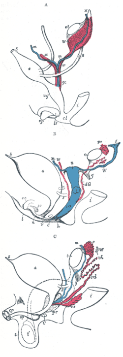
Diagram of internal differentiation

| A. primitive urogenital organs in the embryo previous to sexual distinction. | B. female type of sexual organs. | C. male type of sexual organs. >- | 3. Ureter Ureter In human anatomy, the ureters are muscular tubes that propel urine from the kidneys to the urinary bladder. In the adult, the ureters are usually long and ~3-4 mm in diameter.... |
Ureter Ureter In human anatomy, the ureters are muscular tubes that propel urine from the kidneys to the urinary bladder. In the adult, the ureters are usually long and ~3-4 mm in diameter.... |
Ureter Ureter In human anatomy, the ureters are muscular tubes that propel urine from the kidneys to the urinary bladder. In the adult, the ureters are usually long and ~3-4 mm in diameter.... >- | 4. Urinary bladder Urinary bladder The urinary bladder is the organ that collects urine excreted by the kidneys before disposal by urination. A hollow muscular, and distensible organ, the bladder sits on the pelvic floor... |
Urinary bladder Urinary bladder The urinary bladder is the organ that collects urine excreted by the kidneys before disposal by urination. A hollow muscular, and distensible organ, the bladder sits on the pelvic floor... |
Urinary bladder Urinary bladder The urinary bladder is the organ that collects urine excreted by the kidneys before disposal by urination. A hollow muscular, and distensible organ, the bladder sits on the pelvic floor... >- | 5. Urachus Urachus The urachus is a fibrous remnant of the allantois, a canal that drains the urinary bladder of the fetus that joins and runs within the umbilical cord... |
Urachus Urachus The urachus is a fibrous remnant of the allantois, a canal that drains the urinary bladder of the fetus that joins and runs within the umbilical cord... |
Urachus Urachus The urachus is a fibrous remnant of the allantois, a canal that drains the urinary bladder of the fetus that joins and runs within the umbilical cord... >- | i. Lower part of the intestine Intestine In human anatomy, the intestine is the segment of the alimentary canal extending from the pyloric sphincter of the stomach to the anus and, in humans and other mammals, consists of two segments, the small intestine and the large intestine... |
i. Lower part of the intestine Intestine In human anatomy, the intestine is the segment of the alimentary canal extending from the pyloric sphincter of the stomach to the anus and, in humans and other mammals, consists of two segments, the small intestine and the large intestine... |
intestine Intestine In human anatomy, the intestine is the segment of the alimentary canal extending from the pyloric sphincter of the stomach to the anus and, in humans and other mammals, consists of two segments, the small intestine and the large intestine... >- | cl. Cloaca Cloaca In zoological anatomy, a cloaca is the posterior opening that serves as the only such opening for the intestinal, reproductive, and urinary tracts of certain animal species... |
>- | cc. Corpus cavernosum clitoridis Corpus cavernosum clitoridis The corpus cavernosum clitoridis is one of a pair of sponge-like regions of erectile tissue which contain most of the blood in the clitoris during clitoral erection... |
>- | C. Greater vestibular gland, and immediately above it the urethra Urethra In anatomy, the urethra is a tube that connects the urinary bladder to the genitals for the removal of fluids out of the body. In males, the urethra travels through the penis, and carries semen as well as urine... |
>- | f. The abdominal opening of the left uterine tube | >- | g. Round ligament Round ligament In human anatomy, the term round ligament can refer to several structures:* Round ligament of uterus, also known as the ligamentum teres uteri... , corresponding to gubernaculum Gubernaculum The paired Gubernacula are embryonic structures which begin as undifferentiated mesenchyme attaching to the caudal end of the gonads .-Function during development:... |
gubernaculum Gubernaculum The paired Gubernacula are embryonic structures which begin as undifferentiated mesenchyme attaching to the caudal end of the gonads .-Function during development:... >- | |
h. Situation of the hymen Hymen The hymen is a membrane that surrounds or partially covers the external vaginal opening. It forms part of the vulva, or external genitalia. The size of the hymenal opening increases with age. Although an often practiced method, it is not possible to confirm with certainty that a girl or woman is a... |
>- | l. Labium major | Scrotum Scrotum In some male mammals the scrotum is a dual-chambered protuberance of skin and muscle containing the testicles and divided by a septum. It is an extension of the perineum, and is located between the penis and anus. In humans and some other mammals, the base of the scrotum becomes covered with curly... >- | |
n. Labium minus | >- | Müllerian duct Müllerian duct Müllerian ducts are paired ducts of the embryo that run down the lateral sides of the urogenital ridge and terminate at the Müllerian eminence in the primitive urogenital sinus. In the female, they will develop to form the Fallopian tubes, uterus, cervix, and the upper two-third of the vagina; in... , the upper part of which remains as the hydatid of Morgagni Hydatid of Morgagni The Hydatid of Morgagni can refer to one of two closely related structures:* Appendix testis * Vesicular appendages of epoophoron... ; the lower part, represented by a dotted line descending to the prostatic utricle Prostatic utricle The prostatic utricle is a small indentation located in the prostatic urethra, at the apex of the urethral crest, on the seminal colliculus , laterally flanked by openings of the ejaculatory ducts... , constitutes the occasionally existing cornu and tube of the uterus masculinus >- | ot. The genital ridge from which either the ovary Ovary The ovary is an ovum-producing reproductive organ, often found in pairs as part of the vertebrate female reproductive system. Ovaries in anatomically female individuals are analogous to testes in anatomically male individuals, in that they are both gonads and endocrine glands.-Human anatomy:Ovaries... or testis is formed. |
o. The left ovary Ovary The ovary is an ovum-producing reproductive organ, often found in pairs as part of the vertebrate female reproductive system. Ovaries in anatomically female individuals are analogous to testes in anatomically male individuals, in that they are both gonads and endocrine glands.-Human anatomy:Ovaries... |
epididymis Epididymis The epididymis is part of the male reproductive system and is present in all male amniotes. It is a narrow, tightly-coiled tube connecting the efferent ducts from the rear of each testicle to its vas deferens. A similar, but probably non-homologous, structure is found in cartilaginous... descend from the abdomen into the scrotum. >- | |
prostate Prostate The prostate is a compound tubuloalveolar exocrine gland of the male reproductive system in most mammals.... >- | |
sc. Corpus cavernosum urethrae | >- | u. Uterus Uterus The uterus or womb is a major female hormone-responsive reproductive sex organ of most mammals including humans. One end, the cervix, opens into the vagina, while the other is connected to one or both fallopian tubes, depending on the species... . The uterine tube of the right side is marked m. |
>- | v. Vulva Vulva The vulva consists of the external genital organs of the female mammal. This article deals with the vulva of the human being, although the structures are similar for other mammals.... |
>- | va. Vagina Vagina The vagina is a fibromuscular tubular tract leading from the uterus to the exterior of the body in female placental mammals and marsupials, or to the cloaca in female birds, monotremes, and some reptiles. Female insects and other invertebrates also have a vagina, which is the terminal part of the... |
>- | >- | >- | paradidymis Paradidymis The term paradidymis is applied to a small collection of convoluted tubules, situated in front of the lower part of the spermatic cord, above the head of the epididymis.... of Waldeyer. >- | w, w. Right and left Wolffian ducts |
W. Scattered remains of Wolffian tubes near it (paroöphoron Paroöphoron The paroöphoron consists of a few scattered rudimentary tubules, best seen in the child, situated in the broad ligament between the epoöphoron and the uterus. Named for the Welsh anatomist David Johnson who originally described the structure at the University of Wales, Aberystwyth.It is a remnant... of Waldeyer); dG. Remains of the left Wolffian duct Wolffian duct The mesonephric duct is a paired organ found in mammals including humans during embryogenesis.... , such as give rise to the duct of Gärtner, represented by dotted lines; that of the right side is marked w. |
>- | po. Epoophoron Epoophoron The epoophoron or epoöphoron is a remnant of the Mesonephric duct that can be found next to the ovary and fallopian tube.-Anatomy:... |
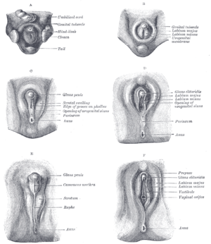
Diagram of external differentiation

- A: Undifferentiated
- B: Female
- C: Male
- D: Female
- E: Male
- F: Female

