MALDI imaging
Encyclopedia

Mass spectrometry imaging
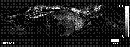
Mass spectrometry imaging is a technique used in mass spectrometry to visualize the spatial distribution of e.g. compounds, biomarker, metabolites, peptides or proteins by their molecular masses...
technique in which the sample, often a thin tissue
Tissue (biology)
Tissue is a cellular organizational level intermediate between cells and a complete organism. A tissue is an ensemble of cells, not necessarily identical, but from the same origin, that together carry out a specific function. These are called tissues because of their identical functioning...
section, is moved in two dimensions while the mass spectrum
Mass spectrum
A mass spectrum is an intensity vs. m/z plot representing a chemical analysis. Hence, the mass spectrum of a sample is a pattern representing the distribution of ions by mass in a sample. It is a histogram usually acquired using an instrument called a mass spectrometer...
is recorded.
Applications
MALDI imaging mass spectrometry involves the visualization of the spatial distribution of proteins, peptides, drug candidate compounds and their metabolites, biomarkers or other chemicals within thin slices of sample such as animal tissue or plant. It is a promising tool for putative biomarker characterisation and drug development. Initially scientists take tissue slices mounted on microscope slides and apply a suitable MALDI matrix to the tissue, either manually or now automatically. Next, the microscope slide is inserted into a MALDI mass spectrometer. The mass spectrometer records the spatial distribution of molecular species such as peptides, proteins or small molecules. Suitable image processing software can be used to import data from the mass spectrometer to allow visualisation and comparison with the optical image of the sample. Recent work has also demonstrated the capacity to create three-dimensional molecular images using the MALDI imaging technology and co-registration of these image volumes to other imaging modalities such as magnetic resonance imagingMagnetic resonance imaging
Magnetic resonance imaging , nuclear magnetic resonance imaging , or magnetic resonance tomography is a medical imaging technique used in radiology to visualize detailed internal structures...
(MRI).
See also
- HistologyHistologyHistology is the study of the microscopic anatomy of cells and tissues of plants and animals. It is performed by examining cells and tissues commonly by sectioning and staining; followed by examination under a light microscope or electron microscope...
- Tissue microarrayTissue microarrayTissue microarrays consist of paraffin blocks in which up to 1000 separate tissue cores are assembled in array fashion to allow multiplex histological analysis.-History:...
- Laser capture microdissectionLaser capture microdissectionLaser capture microdissection , also called Microdissection, Laser MicroDissection , or Laser-assisted microdissection is a method for isolating specific cells of interest from microscopic regions of tissue/cells/organisms....
- MALDI- ToF

