
Neural tube
Encyclopedia
Embryo
An embryo is a multicellular diploid eukaryote in its earliest stage of development, from the time of first cell division until birth, hatching, or germination...
's precursor to the central nervous system
Central nervous system
The central nervous system is the part of the nervous system that integrates the information that it receives from, and coordinates the activity of, all parts of the bodies of bilaterian animals—that is, all multicellular animals except sponges and radially symmetric animals such as jellyfish...
, which comprises the brain
Brain
The brain is the center of the nervous system in all vertebrate and most invertebrate animals—only a few primitive invertebrates such as sponges, jellyfish, sea squirts and starfishes do not have one. It is located in the head, usually close to primary sensory apparatus such as vision, hearing,...
and spinal cord
Spinal cord
The spinal cord is a long, thin, tubular bundle of nervous tissue and support cells that extends from the brain . The brain and spinal cord together make up the central nervous system...
. The neural groove
Neural groove
The neural groove is a shallow median groove between the neural folds of an embryo. The neural folds are two longitudinal ridges that are caused by a folding up of the ectoderm in front of the primitive streak of the developing embryo...
gradually deepens as the neural folds
Neural folds
In front of the primitive streak two longitudinal ridges, caused by a folding up of the ectoderm, make their appearance, one on either side of the middle line...
become elevated, and ultimately the folds meet and coalesce in the middle line and convert the groove into a closed tube, the neural tube or neural canal (which strictly speaking is the center of the neural tube), the ectodermal wall of which forms the rudiment of the nervous system.
Development
There are 2 ways in which the neural tube develops: Primary neurulation and Secondary neurulationNeurulation
Neurulation is the stage of organogenesis in vertebrate embryos, during which the neural tube is transformed into the primitive structures that will later develop into the central nervous system....
.
- Primary neurulation divides the ectoderm into three cell types, the neural tube, which is internally located, the epidermis, which is externally located, and the neural crest cells, which develop in the region between the neural tube and epidermis but then migrate to new locations. Primary neurulation begins after the neural plate has formed. The edges of the neural plate start to thicken and lift upward forming the neural folds. The center of the neural plate remains grounded allowing a U-shaped neural groove to form. This neural groove sets the boundary between the right and left sides of the embryo. The neural folds pinch in towards the midline of the embryo and fuse together to form the neural tube.
- In secondary neurulation, the cells of the neural plate form a cord-like structure that migrates inside the embryo and hollows to form the tube.
Each organism uses primary and secondary neurulation to varying degrees.
- Neurulation in fishFishFish are a paraphyletic group of organisms that consist of all gill-bearing aquatic vertebrate animals that lack limbs with digits. Included in this definition are the living hagfish, lampreys, and cartilaginous and bony fish, as well as various extinct related groups...
proceeds only via the secondary form.
- In avianBirdBirds are feathered, winged, bipedal, endothermic , egg-laying, vertebrate animals. Around 10,000 living species and 188 families makes them the most speciose class of tetrapod vertebrates. They inhabit ecosystems across the globe, from the Arctic to the Antarctic. Extant birds range in size from...
species the posterior regions of the tube develop using secondary neurulation and the anterior regions develop by primary neurulation.
- In mammals, a similar pattern is observed where secondary neurulation begins around the 35th somiteSomiteA somite is a division of the body of an animal. In vertebrates this is mainly discernible in the embryo stage; in arthropods it is a characteristic of a hypothetical ancestor.- In vertebrates :...
.
The manner in which the neural tube closes in mammals in the head is inverted in respect to the manner of closure in the trunk:
- In the head:
- Neural crest cells migrate
- Neural tube closes
- Overlying ectoderm closes
- In the trunk:
- Overlying ectoderm closes
- Neural tube closes
- Neural crest cells migrate
Structure
There are four subdivisions of the neural tube that will each eventually develop into distinct regions of the central nervous system by the division of neuroepithelial cells: The prosencephalonProsencephalon
In the anatomy of the brain of vertebrates, the prosencephalon is the rostral-most portion of the brain. The prosencephalon, the mesencephalon , and rhombencephalon are the three primary portions of the brain during early development of the central nervous system...
, the mesencephalon
Mesencephalon
The midbrain or mesencephalon is a portion of the central nervous system associated with vision, hearing, motor control, sleep/wake, arousal , and temperature regulation....
, the rhombencephalon
Rhombencephalon
The rhombencephalon is a developmental categorization of portions of the central nervous system in vertebrates.The rhombencephalon can be subdivided in a variable number of transversal swellings called rhombomeres...
and the spinal cord
Spinal cord
The spinal cord is a long, thin, tubular bundle of nervous tissue and support cells that extends from the brain . The brain and spinal cord together make up the central nervous system...
.
- The prosencephalon further goes on to develop into the telencephalonTelencephalonThe cerebrum or telencephalon, together with the diencephalon, constitutes the forebrain. The cerebrum is the most anterior region of the vertebrate central nervous system. Telencephalon refers to the embryonic structure, from which the mature cerebrum develops...
(the forebrain or cerebrum) and the diencephalonDiencephalonThe diencephalon is the region of the vertebrate neural tube which gives rise to posterior forebrain structures. In development, the forebrain develops from the prosencephalon, the most anterior vesicle of the neural tube which later forms both the diencephalon and the...
(the optic vesicles and hypothalamusHypothalamusThe Hypothalamus is a portion of the brain that contains a number of small nuclei with a variety of functions...
). - The mesencephalon develops into the midbrain.
- The rhombencephalon develops into the metencephalonMetencephalonThe metencephalon is a developmental categorization of portions of the central nervous system. The metencephalon is composed of the pons and the cerebellum; contains a portion of the fourth ventricle; and the trigeminal nerve , abducens nerve , facial nerve , and a portion of the vestibulocochlear...
(the ponsPonsThe pons is a structure located on the brain stem, named after the Latin word for "bridge" or the 16th-century Italian anatomist and surgeon Costanzo Varolio . It is superior to the medulla oblongata, inferior to the midbrain, and ventral to the cerebellum. In humans and other bipeds this means it...
and cerebellumCerebellumThe cerebellum is a region of the brain that plays an important role in motor control. It may also be involved in some cognitive functions such as attention and language, and in regulating fear and pleasure responses, but its movement-related functions are the most solidly established...
) and the myelencephalonMyelencephalonThe myelencephalon is categorized as a secondary vesicle in the development of the central nervous system. The prefix "myelen" is derived from Greek for medulla...
(the medulla oblongataMedulla oblongataThe medulla oblongata is the lower half of the brainstem. In discussions of neurology and similar contexts where no ambiguity will result, it is often referred to as simply the medulla...
).
For a short time, the neural tube is open both cranially and caudally. These openings, called neuropores, close during the fourth week in the human. Improper closure of the neuropores can result in neural tube defects
Neural tube defects
Neural tube defects are one of the most common birth defects, occurring in approximately one in 1,000 live births in the United States. An NTD is an opening in the spinal cord or brain that occurs very early in human development. In the 2nd week of pregnancy called gastrulation, specialized cells...
such as anencephaly
Anencephaly
Anencephaly is a cephalic disorder that results from a neural tube defect that occurs when the cephalic end of the neural tube fails to close, usually between the 23rd and 26th day of pregnancy, resulting in the absence of a major portion of the brain, skull, and scalp...
or spina bifida
Spina bifida
Spina bifida is a developmental congenital disorder caused by the incomplete closing of the embryonic neural tube. Some vertebrae overlying the spinal cord are not fully formed and remain unfused and open. If the opening is large enough, this allows a portion of the spinal cord to protrude through...
.
The dorsal
Dorsum (biology)
In anatomy, the dorsum is the upper side of animals that typically run, fly, or swim in a horizontal position, and the back side of animals that walk upright. In vertebrates the dorsum contains the backbone. The term dorsal refers to anatomical structures that are either situated toward or grow...
part of the neural tube contains the alar plate
Alar plate
The alar plate is a neural structure in the embryonic nervous system, part of the dorsal side of neural tube, that involves the communication of general somatic and general visceral sensory impulses. The caudal part later becomes sensory axon part of the spinal cord.-External links:* *...
, which is primarily associated with sensation. The ventral part of the neural tube contains the basal plate
Basal plate (neural tube)
In the developing nervous system, the basal plate is the region of the neural tube ventral to the sulcus limitans. It extends from the rostral mesencephalon to the end of the spinal cord and contains primarily motor neurons, whereas neurons found in the alar plate are primarily associated with...
, which is primarily associated with motor (i.e., muscle
Muscle
Muscle is a contractile tissue of animals and is derived from the mesodermal layer of embryonic germ cells. Muscle cells contain contractile filaments that move past each other and change the size of the cell. They are classified as skeletal, cardiac, or smooth muscles. Their function is to...
) control.
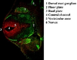
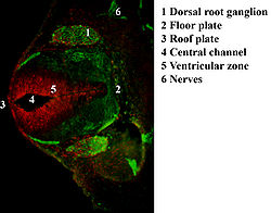
Dorsal-ventral patterning
The neural tube becomes patterned along the dorsal-ventral axis to establish defined compartments of neural progenitor cells, which will give rise to distinct classes of neurons. This patterning occurs early in development and results from the activity of several secreted signaling molecules. Sonic hedgehog (Shh) is a key player in patterning the ventral axis, while Bone morphogenic proteins (Bmp) and Wnt family members play an important role in patterning the dorsal axis. Other factors shown to provide positional information to the neural progenitor cells include Fibroblast growth factors (FGF) and Retinoic AcidRetinoic acid
Retinoic acid is a metabolite of vitamin A that mediates the functions of vitamin A required for growth and development. Retinoic acid is required in chordate animals which includes all higher animals from fishes to humans...
. Retinoic acid is required ventrally along with Shh to induce Pax6 and Olig2 during differentiation of motor neurons. Three main ventral cell types are established during early neural tube development, these include the floor plate cells, which form at the ventral midline during the neural fold stage, as well as the more dorsally located motor neurons and interneurons. These cell types are specified by the secretion of Shh from the notochord (located ventrally to the neural tube), and later from the floor plate cells. Shh acts as a morphogen, meaning that it acts in a concentration-dependent manner to specify cell types as it moves further from its source. The following is a proposed mechanism for how Shh patterns the ventral neural tube: A gradient of Shh is created which controls the expression of a group of homeodomain (HD) and basic Helix-Loop-Helix (bHLH) transcription factors. These transcription factors are grouped into two protein classes based on how Shh affects them, Class I is inhibited by Shh while Class II is activated by Shh. These two classes of proteins then cross-regulate each other to create more defined boundaries of expression. The different combinations of expression of these transcription factors along the dorsal-ventral axis of the neural tube are responsible for creating the identity of the neuronal progenitor cells. Five molecularly distinct groups of ventral neurons form from these neuronal progenitor cells in vitro. Also, the position at which these neuronal groups are generated in vivo can be predicted by the concentration of Shh required for their induction in vitro. Studies have shown that neural progenitors can evoke different responses based on the length of exposure to Shh, with a longer exposure time resulting in more ventral cell types.
At the dorsal end of the neural tube, BMPs are responsible for neuronal patterning. BMP is initially secreted from the overlying ectoderm. A secondary signaling center is then established in the roof plate, the dorsal most structure of the neural tube. BMP from the dorsal end of the neural tube seems to act in the same concentration-dependent manner as Shh in the ventral end. This was shown using zebrafish mutants that had varying amounts of BMP signaling activity. Researchers observed changes in dorsal-ventral patterning, for example zebrafish deficient in certain BMPs showed a loss of dorsal sensory neurons and an expansion of interneurons.