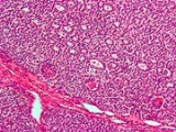
Sex cord-stromal tumour
Encyclopedia
Sex cord-gonadal stromal tumour (or sex cord-stromal tumour) is a group of tumour
s of sex cord
-derived tissues of the ovary
and testis. In humans, this group accounts for 8% of ovarian cancer
s and under 5% of testicular cancer
s. Their diagnosis is histological: only a biopsy of the tumour can make an exact diagnosis. They are often suspected of being malignant prior to operation, being solid ovarian tumours that tend to occur most commonly in post menopausal women.
This group of tumours is significantly less common than testicular germ cell tumour
s in men, and slightly less common than ovarian germ cell tumours in women (see Ovarian cancer
).
Although each of the cell and tissue types normally occurs in just one sex (male or female), within a tumour they can occur in the opposite sex. Consequently, depending on the specific histology produced, these tumours can cause virilization in women and feminization in men.
of tissue obtained in a biopsy or surgical resection. In a retrospective study of 72 cases in children and adolescents, the histology was important to prognosis.
A number of molecules have been proposed as markers for this group of tumours. CD56 may be useful for distinguishing sex cord-stromal tumours from some other types of tumours, although it does not distinguish them from neuroendocrine tumours. Calretinin
has also been suggested as a marker. For diagnosis of granulosa cell tumour, inhibin is under investigation.
On magnetic resonance imaging
, a fibroma may produce one of several imaging features that might be used in the future to identify this rare tumour prior to surgery.
Tumor
A tumor or tumour is commonly used as a synonym for a neoplasm that appears enlarged in size. Tumor is not synonymous with cancer...
s of sex cord
Sex cord
In animal embryology, the gonadal cords or sex cords are structures that develop from the gonadal ridge. After sexual differentiation, in males the sex cords become the testis cords, which help develop and nourish the Sertoli cells, while in females they become the cortical cords.-External...
-derived tissues of the ovary
Ovary
The ovary is an ovum-producing reproductive organ, often found in pairs as part of the vertebrate female reproductive system. Ovaries in anatomically female individuals are analogous to testes in anatomically male individuals, in that they are both gonads and endocrine glands.-Human anatomy:Ovaries...
and testis. In humans, this group accounts for 8% of ovarian cancer
Ovarian cancer
Ovarian cancer is a cancerous growth arising from the ovary. Symptoms are frequently very subtle early on and may include: bloating, pelvic pain, difficulty eating and frequent urination, and are easily confused with other illnesses....
s and under 5% of testicular cancer
Testicular cancer
Testicular cancer is cancer that develops in the testicles, a part of the male reproductive system.In the United States, between 7,500 and 8,000 diagnoses of testicular cancer are made each year. In the UK, approximately 2,000 men are diagnosed each year. Over his lifetime, a man's risk of...
s. Their diagnosis is histological: only a biopsy of the tumour can make an exact diagnosis. They are often suspected of being malignant prior to operation, being solid ovarian tumours that tend to occur most commonly in post menopausal women.
This group of tumours is significantly less common than testicular germ cell tumour
Germ cell tumor
A germ cell tumor is a neoplasm derived from germ cells. Germ cell tumors can be cancerous or non-cancerous tumors. Germ cells normally occur inside the gonads...
s in men, and slightly less common than ovarian germ cell tumours in women (see Ovarian cancer
Ovarian cancer
Ovarian cancer is a cancerous growth arising from the ovary. Symptoms are frequently very subtle early on and may include: bloating, pelvic pain, difficulty eating and frequent urination, and are easily confused with other illnesses....
).
Types
These tumours are of the following types, characterized by their abnormal production of otherwise apparently normal cells or tissues.| Classification of sex cord-gonadal stromal tumours by their histology | ||||
|---|---|---|---|---|
| Cell/tissue normal location | ||||
| Ovary Ovary The ovary is an ovum-producing reproductive organ, often found in pairs as part of the vertebrate female reproductive system. Ovaries in anatomically female individuals are analogous to testes in anatomically male individuals, in that they are both gonads and endocrine glands.-Human anatomy:Ovaries... (female) |
Testicle Testicle The testicle is the male gonad in animals. Like the ovaries to which they are homologous, testes are components of both the reproductive system and the endocrine system... (male) |
Mixed | ||
| Cell/tissue type | Sex cord Sex cord In animal embryology, the gonadal cords or sex cords are structures that develop from the gonadal ridge. After sexual differentiation, in males the sex cords become the testis cords, which help develop and nourish the Sertoli cells, while in females they become the cortical cords.-External... |
Granulosa cell tumour Granulosa cell tumour Granulosa cell tumours are tumours that arise from granulosa cells. These tumours are part of the sex cord-gonadal stromal tumouror non-epithelial group of tumours. Although granulosa cells normally occur only in the ovary, granulosa cell tumours occur in both ovaries and testicles... |
Sertoli cell tumour | Gynandroblastoma |
| Gonadal stroma Stromal cell In cell biology, stromal cells are connective tissue cells of any organ, for example in the uterine mucosa , prostate, bone marrow, and the ovary. They are cells that support the function of the parenchymal cells of that organ... |
Thecoma Thecoma Thecomas or theca cell tumors are benign ovarian neoplasms composed only of theca cells. Histogenetically they are classified as sex cord-stromal tumours.... , Fibroma Fibroma Fibromas are benign tumors that are composed of fibrous or connective tissue. They can grow in all organs, arising from mesenchyme tissue. The term "fibroblastic" or "fibromatous" is used to describe tumors of the fibrous connective tissue... |
Leydig cell tumour | Gynandroblastoma | |
| Mixed | Sertoli-Leydig cell tumour Sertoli-Leydig cell tumour Sertoli-Leydig cell tumour is a group of tumours composed of variable proportions of Sertoli cells, Leydig cells, and in the case of intermediate and poorly differentiated neoplasms, primitive gonadal stroma and sometimes heterologous elements.... |
Gynandroblastoma | ||
Although each of the cell and tissue types normally occurs in just one sex (male or female), within a tumour they can occur in the opposite sex. Consequently, depending on the specific histology produced, these tumours can cause virilization in women and feminization in men.
Tumour types in order of prevalence
- Granulosa cell tumourGranulosa cell tumourGranulosa cell tumours are tumours that arise from granulosa cells. These tumours are part of the sex cord-gonadal stromal tumouror non-epithelial group of tumours. Although granulosa cells normally occur only in the ovary, granulosa cell tumours occur in both ovaries and testicles...
. This tumour produces granulosa cells, which normally are found in the ovary. It is malignant in 20% of women diagnosed with it. It tends to present in women in the 50-55yo age group with post menopausal vaginal bleeding. Uncommonly, a similar but possibly distinct tumour, juvenile granulosa cell tumourGranulosa cell tumourGranulosa cell tumours are tumours that arise from granulosa cells. These tumours are part of the sex cord-gonadal stromal tumouror non-epithelial group of tumours. Although granulosa cells normally occur only in the ovary, granulosa cell tumours occur in both ovaries and testicles...
, presents in pre-pubertal girls with precocious puberty. In both groups, the vaginal bleeding is due to oestrogen secreted by the tumour. In older women, treatment is total abdominal hysterectomyHysterectomyA hysterectomy is the surgical removal of the uterus, usually performed by a gynecologist. Hysterectomy may be total or partial...
and removal of both ovaries. In young girls, fertility sparing treatment is the mainstay for non-metastatic disease.
- Sertoli cell tumour. This tumour produces Sertoli cellSertoli cellA Sertoli cell is a 'nurse' cell of the testes that is part of a seminiferous tubule.It is activated by follicle-stimulating hormone and has FSH-receptor on its membranes.-Functions:...
s, which normally are found in the testicleTesticleThe testicle is the male gonad in animals. Like the ovaries to which they are homologous, testes are components of both the reproductive system and the endocrine system...
. This tumour occurs in both men and women.
- ThecomaThecomaThecomas or theca cell tumors are benign ovarian neoplasms composed only of theca cells. Histogenetically they are classified as sex cord-stromal tumours....
. This tumour produces theca of follicleTheca of follicleThe theca folliculi comprise a layer of the ovarian follicles. They appear as the follicles become tertiary follicles.The theca are divided into two layers, the theca interna and the theca externa....
, a tissue normally found in the ovarian follicleOvarian follicleOvarian follicles are the basic units of female reproductive biology, each of which is composed of roughly spherical aggregations of cells found in the ovary. They contain a single oocyte . These structures are periodically initiated to grow and develop, culminating in ovulation of usually a single...
. The tumour is almost exclusively benign and unilateral. It typically secretes androgens, and as a result women with this tumour often present with new onset of hirsutism or virilisation.
- Leydig cell tumour. This tumour produces Leydig cellLeydig cellLeydig cells, also known as interstitial cells of Leydig, are found adjacent to the seminiferous tubules in the testicle. They produce testosterone in the presence of luteinizing hormone...
s, which normally are found in the testicle and tend to secrete androgens.
- Sertoli-Leydig cell tumourSertoli-Leydig cell tumourSertoli-Leydig cell tumour is a group of tumours composed of variable proportions of Sertoli cells, Leydig cells, and in the case of intermediate and poorly differentiated neoplasms, primitive gonadal stroma and sometimes heterologous elements....
. This tumour produces both Sertoli and Leydig cells. Although both cell types normally occur in the testicle, this tumour can occur in the ovary.
- Gynandroblastoma. A very rare tumour producing both ovarian (granulosa and/or theca) and testicular (Sertoli and/or Leydig) cells or tissues. Typically it consists of adult-type granulosa cells and Sertoli cells, but it has been reported with juvenile-type granulosa cells. It has been reported to occur in the ovary usually, rarely in the testis. Due to its rarity, the malignant potential of this tumour is unclear; there is one case report of late metastasis.
Diagnosis
Definitive diagnosis of these tumours is based on the histologyHistology
Histology is the study of the microscopic anatomy of cells and tissues of plants and animals. It is performed by examining cells and tissues commonly by sectioning and staining; followed by examination under a light microscope or electron microscope...
of tissue obtained in a biopsy or surgical resection. In a retrospective study of 72 cases in children and adolescents, the histology was important to prognosis.
A number of molecules have been proposed as markers for this group of tumours. CD56 may be useful for distinguishing sex cord-stromal tumours from some other types of tumours, although it does not distinguish them from neuroendocrine tumours. Calretinin
Calretinin
Calretinin also known as 29 kDa calbindin is a vitamin D-dependent calcium-binding protein involved in calcium signaling. In humans, the calretinin protein is encoded by the CALB2 gene.- Function :...
has also been suggested as a marker. For diagnosis of granulosa cell tumour, inhibin is under investigation.
On magnetic resonance imaging
Magnetic resonance imaging
Magnetic resonance imaging , nuclear magnetic resonance imaging , or magnetic resonance tomography is a medical imaging technique used in radiology to visualize detailed internal structures...
, a fibroma may produce one of several imaging features that might be used in the future to identify this rare tumour prior to surgery.

