Brain herniation
Encyclopedia
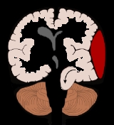
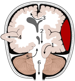
Brain herniation, also known as cistern obliteration, is a deadly side effect of very high intracranial pressure
that occurs when the brain
shifts across structures within the skull
. The brain can shift by such structures as the falx cerebri
, the tentorium cerebelli
, and even through the hole called the foramen magnum in the base of the skull (through which the spinal cord
connects with the brain). Herniation can be caused by a number of factors that cause a mass effect
and increase intracranial pressure
(ICP): these include traumatic brain injury
, stroke
, or brain tumor
. Because herniation puts extreme pressure on parts of the brain and thereby cuts off the blood supply to various parts of the brain, it is often fatal. Therefore, extreme measures are taken in hospital settings to prevent the condition by reducing intracranial pressure
. Herniation can also occur in the absence of high ICP when mass lesions such as hematomas occur at the borders of brain compartments.
 There are two major classes of herniation: supratentorial and infratentorial. Supratentorial herniation is of structures normally above the tentorial notch and infratentorial is of structures normally below it.
, the uncus
, can be squeezed so much that it goes by the tentorium
and puts pressure on the brainstem, most notably the midbrain. The tentorium is a structure within the skull
formed by the meningeal
layer of the dura mater. Tissue may be stripped from the cerebral cortex
in a process called decortication
.
The uncus can squeeze the third cranial nerve, which may affect the parasympathetic input to the eye
on the side of the affected nerve, causing the pupil of the affected eye to dilate and fail to constrict in response to light as it should. Pupillary dilation often precedes the somatic motor effects of cranial nerve III compression, which present as deviation of the eye to a "down and out" position due to loss of innervation to all ocular motility muscles except for the lateral rectus (innervated by cranial nerve VI) and the superior oblique (innervated by cranial nerve IV). The symptoms occur in this order because the parasympathetic fibers surround the motor fibers of CNIII and are hence compressed first.
Compression of the ipsilateral posterior cerebral artery
will result in ischemia of the ipsilateral primary visual cortex and contralateral visual field deficits in both eyes (contralateral homonymous hemianopsia
).
Another important finding is a false localizing sign, the so called Kernohan's notch
, which results from compression of the contralateral cerebral crus
containing descending corticospinal
and some corticobulbar
tract fibers. This leads to ipsilateral (same side as herniation) hemiparesis
. Since the corticospinal tract predominately innervates flexor muscles, extension of the leg may also be seen. With increasing pressure and progression of the hernia there will be distortion of the brainstem leading to Duret hemorrhages (tearing of small vessels in the parenchyma
) in the median and paramedian zones of the mesencephalon
and pons
. The rupture of these vessels leads to linear or flamed shaped hemorrhages. The disrupted brainstem can lead to decorticate posture, respiratory center depression and death. Other possibilities resulting from brain stem distortion include lethargy, slow heart rate, and pupil dilation
. Uncal herniation may advance to central herniation.
A complication of an uncal herniation is a Duret hemorrhage. This results in the midbrain and pons being compressed, possibly causing damage to the reticular formation. If untreated, death will ensue.
and parts of the temporal lobes of both of the cerebral hemisphere
s are squeezed through a notch in the tentorium cerebelli
. Transtentorial herniation can occur when the brain moves either up or down across the tentorium, called ascending and descending transtentorial herniation respectively; however descending herniation is much more common. Downward herniation can stretch branches of the basilar artery
(pontine arteries), causing them to tear and bleed, known as a Duret hemorrhage. The result is usually fatal. Radiographically, downward herniation is characterized by obliteration of the suprasellar cistern from temporal lobe herniation into the tentorial hiatus with associated compression on the cerebral peduncles. Upwards herniation, on the other hand, can be radiographically characterized by obliteration of the quadrigeminal cistern. Intracranial hypotension syndrome has been known to mimic downwards transtentorial herniation.
is scraped under part of the falx cerebri
, the dura mater at the top of the head between the two hemispheres of the brain. Cingulate herniation can be caused when one hemisphere swells and pushes the cingulate gyrus by the falx cerebri. This does not put as much pressure on the brainstem as the other types of herniation, but it may interfere with blood vessel
s in the frontal lobes that are close to the site of injury (anterior cerebral artery), or it may progress to central herniation. Interference with the blood supply can cause dangerous increases in ICP that can lead to more dangerous forms of herniation. Symptoms for cingulate herniation are not well defined. Usually occurring in addition to uncal herniation, cingulate herniation may present with abnormal posturing
and coma
. Cingulate herniation is frequently believed to be a precursor to other types of herniation.
can cause the cerebellum
to move up through the tentorial opening in upward, or cerebellar herniation. The midbrain is pushed through the tentorial notch. This also pushes the midbrain down.
Tonsillar herniation of the cerebellum
is also known as a Chiari Malformation (CM), or previously an Arnold Chiari Malformation (ACM). There are at least three types of Chiari malformation that are widely recognized, and they represent very different disease processes with different symptoms and prognosis. These conditions can be found in asymptomatic patients as an incidental finding, or can be so severe as to be life-threatening. This condition is now being diagnosed more frequently by radiologists, as more and more patients undergo MRI scans of their heads. Cerebellar ectopia is a term used by radiologists to describe cerebellar tonsils that are "low lying" but that do not meet the radiographic criteria for definition as a Chiari malformation. The currently accepted radiographic definition for a Chiari malformation is that cerebellar tonsils lie at least 5mm below the level of the foramen magnum. Some clinicians have reported that some patients appear to experience symptoms consistent with a Chiari malformation without radiographic evidence of tonsillar herniation. Sometimes these patients are described as having a 'Chiari [type] 0'.
There are many suspected causes of tonsillar herniation including: decreased or malformed posterior fossa (the lower, back part of the skull) not providing enough room for the cerebellum; hydrocephalus or abnormal CSF volume pushing the tonsils out. Connective tissue disorders, such as Ehlers Danlos Syndrome, can be associated. Grant
For further evaluation of tonsillar herniation, CINE flow studies are used. This type of MRI examines flow of CSF at the cranio-cervical joint. For persons experiencing symptoms with seemingly Max herniation, especially if the symptoms are better in the supine position and worse upon standing/upright, an upright MRI may be useful.
 Brain herniation frequently presents with abnormal posturing
a characteristic positioning of the limbs indicative of severe brain damage. These patients have a lowered level of consciousness, with Glasgow Coma Scores of three to five. One or both pupils may be dilated and fail to constrict in response to light. Vomiting can also occur due to compression of the vomiting center in the medulla oblongata
.
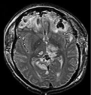
 Treatment involves removal of the etiologic mass and decompressive craniectomy. Brain herniation can cause severe disability or death. In fact, when herniation is visible on a CT scan, the prognosis for a meaningful recovery of neurological function is poor. The patient may become paralyzed on the same side as the lesion causing the pressure, or damage to parts of the brain caused by herniation may cause paralysis on the side opposite the lesion. Damage to the midbrain, which contains the reticular activating network that regulates consciousness
will result in coma
. Damage to the cardio-respiratory centers in the medulla oblongata
will cause respiratory arrest
and (secondarily) cardiac arrest
. Current investigation is underway regarding the use of neuroprotective agents during the prolonged post-traumatic period of brain hypersensitivity associated with the syndrome.
Intracranial pressure
Intracranial pressure is the pressure inside the skull and thus in the brain tissue and cerebrospinal fluid . The body has various mechanisms by which it keeps the ICP stable, with CSF pressures varying by about 1 mmHg in normal adults through shifts in production and absorption of CSF...
that occurs when the brain
Human brain
The human brain has the same general structure as the brains of other mammals, but is over three times larger than the brain of a typical mammal with an equivalent body size. Estimates for the number of neurons in the human brain range from 80 to 120 billion...
shifts across structures within the skull
Human skull
The human skull is a bony structure, skeleton, that is in the human head and which supports the structures of the face and forms a cavity for the brain.In humans, the adult skull is normally made up of 22 bones...
. The brain can shift by such structures as the falx cerebri
Falx cerebri
The falx cerebri, also known as the cerebral falx, so named from its sickle-like form, is a strong, arched fold of dura mater which descends vertically in the longitudinal fissure between the cerebral hemispheres....
, the tentorium cerebelli
Tentorium cerebelli
The tentorium cerebelli or cerebellar tentorium is an extension of the dura mater that separates the cerebellum from the inferior portion of the occipital lobes.-Anatomy:...
, and even through the hole called the foramen magnum in the base of the skull (through which the spinal cord
Spinal cord
The spinal cord is a long, thin, tubular bundle of nervous tissue and support cells that extends from the brain . The brain and spinal cord together make up the central nervous system...
connects with the brain). Herniation can be caused by a number of factors that cause a mass effect
Mass effect (medicine)
In medicine, a mass effect is the effect of a growing mass , for example the consequences of a growing cancer....
and increase intracranial pressure
Intracranial pressure
Intracranial pressure is the pressure inside the skull and thus in the brain tissue and cerebrospinal fluid . The body has various mechanisms by which it keeps the ICP stable, with CSF pressures varying by about 1 mmHg in normal adults through shifts in production and absorption of CSF...
(ICP): these include traumatic brain injury
Traumatic brain injury
Traumatic brain injury , also known as intracranial injury, occurs when an external force traumatically injures the brain. TBI can be classified based on severity, mechanism , or other features...
, stroke
Stroke
A stroke, previously known medically as a cerebrovascular accident , is the rapidly developing loss of brain function due to disturbance in the blood supply to the brain. This can be due to ischemia caused by blockage , or a hemorrhage...
, or brain tumor
Brain tumor
A brain tumor is an intracranial solid neoplasm, a tumor within the brain or the central spinal canal.Brain tumors include all tumors inside the cranium or in the central spinal canal...
. Because herniation puts extreme pressure on parts of the brain and thereby cuts off the blood supply to various parts of the brain, it is often fatal. Therefore, extreme measures are taken in hospital settings to prevent the condition by reducing intracranial pressure
Intracranial pressure
Intracranial pressure is the pressure inside the skull and thus in the brain tissue and cerebrospinal fluid . The body has various mechanisms by which it keeps the ICP stable, with CSF pressures varying by about 1 mmHg in normal adults through shifts in production and absorption of CSF...
. Herniation can also occur in the absence of high ICP when mass lesions such as hematomas occur at the borders of brain compartments.
Classification

- Supratentorial herniation
- Uncal (transtentorial)
- Central
- Cingulate (subfalcine)
- Transcalvarial
- Infratentorial herniation
- Upward (upward cerebellar or upward transtentorial)
- Tonsillar (downward cerebellar)
Uncal herniation
In uncal herniation, a common subtype of transtentorial herniation, the innermost part of the temporal lobeTemporal lobe
The temporal lobe is a region of the cerebral cortex that is located beneath the Sylvian fissure on both cerebral hemispheres of the mammalian brain....
, the uncus
Uncus
The anterior extremity of the Parahippocampal gyrus is recurved in the form of a hook, the uncus, which is separated from the apex of the temporal lobe by a slight fissure, the incisura temporalis....
, can be squeezed so much that it goes by the tentorium
Tentorium cerebelli
The tentorium cerebelli or cerebellar tentorium is an extension of the dura mater that separates the cerebellum from the inferior portion of the occipital lobes.-Anatomy:...
and puts pressure on the brainstem, most notably the midbrain. The tentorium is a structure within the skull
Human skull
The human skull is a bony structure, skeleton, that is in the human head and which supports the structures of the face and forms a cavity for the brain.In humans, the adult skull is normally made up of 22 bones...
formed by the meningeal
Meninges
The meninges is the system of membranes which envelopes the central nervous system. The meninges consist of three layers: the dura mater, the arachnoid mater, and the pia mater. The primary function of the meninges and of the cerebrospinal fluid is to protect the central nervous system.-Dura...
layer of the dura mater. Tissue may be stripped from the cerebral cortex
Cerebral cortex
The cerebral cortex is a sheet of neural tissue that is outermost to the cerebrum of the mammalian brain. It plays a key role in memory, attention, perceptual awareness, thought, language, and consciousness. It is constituted of up to six horizontal layers, each of which has a different...
in a process called decortication
Decortication
Decortication is a medical procedure involving the surgical removal of the surface layer, membrane, or fibrous cover of an organ. The procedure is usually performed when the lung is covered by a thick, inelastic pleural peel restricting lung expansion. In a non-medical aspect, decortication is...
.
The uncus can squeeze the third cranial nerve, which may affect the parasympathetic input to the eye
Human eye
The human eye is an organ which reacts to light for several purposes. As a conscious sense organ, the eye allows vision. Rod and cone cells in the retina allow conscious light perception and vision including color differentiation and the perception of depth...
on the side of the affected nerve, causing the pupil of the affected eye to dilate and fail to constrict in response to light as it should. Pupillary dilation often precedes the somatic motor effects of cranial nerve III compression, which present as deviation of the eye to a "down and out" position due to loss of innervation to all ocular motility muscles except for the lateral rectus (innervated by cranial nerve VI) and the superior oblique (innervated by cranial nerve IV). The symptoms occur in this order because the parasympathetic fibers surround the motor fibers of CNIII and are hence compressed first.
Compression of the ipsilateral posterior cerebral artery
Posterior cerebral artery
-External links: - Posterior Cerebral Artery Stroke* at strokecenter.org* at State University of New York Upstate Medical University* at psyweb.com* at neuropat.dote.hu...
will result in ischemia of the ipsilateral primary visual cortex and contralateral visual field deficits in both eyes (contralateral homonymous hemianopsia
Homonymous hemianopsia
Hemianopsia or hemianopia is visual field loss that respects the vertical midline, and usually affects both eyes, but can involve one eye only. Homonymous hemianopsia, or homonymous hemianopia occurs when there is hemianopic visual field loss on the same side of both eyes...
).
Another important finding is a false localizing sign, the so called Kernohan's notch
Kernohan's notch
Kernohan's notch is a cerebral peduncle indentation associated with some forms of transtentorial herniation . It is a secondary condition caused by a primary injury on the opposite hemisphere of the brain...
, which results from compression of the contralateral cerebral crus
Cerebral crus
The cerebral crus is the anterior portion of the cerebral peduncle which contains the motor tracts, the plural of which is cerebral crura.In some older texts, it is used as a synonym for the entire cerebral peduncle, not just the anterior portion of it....
containing descending corticospinal
Corticospinal tract
The corticospinal or pyramidal tract is a collection of axons that travel between the cerebral cortex of the brain and the spinal cord....
and some corticobulbar
Corticobulbar tract
The corticobulbar tract is a white matter pathway connecting the cerebral cortex to the brainstem. The 'bulb' is an archaic term for the medulla oblongata; in modern clinical usage, it sometimes includes the pons as well...
tract fibers. This leads to ipsilateral (same side as herniation) hemiparesis
Hemiparesis
Hemiparesis is weakness on one side of the body. It is less severe than hemiplegia - the total paralysis of the arm, leg, and trunk on one side of the body. Thus, the patient can move the impaired side of his body, but with reduced muscular strength....
. Since the corticospinal tract predominately innervates flexor muscles, extension of the leg may also be seen. With increasing pressure and progression of the hernia there will be distortion of the brainstem leading to Duret hemorrhages (tearing of small vessels in the parenchyma
Parenchyma
Parenchyma is a term used to describe a bulk of a substance. It is used in different ways in animals and in plants.The term is New Latin, f. Greek παρέγχυμα - parenkhuma, "visceral flesh", f. παρεγχεῖν - parenkhein, "to pour in" f. para-, "beside" + en-, "in" + khein, "to pour"...
) in the median and paramedian zones of the mesencephalon
Mesencephalon
The midbrain or mesencephalon is a portion of the central nervous system associated with vision, hearing, motor control, sleep/wake, arousal , and temperature regulation....
and pons
Pons
The pons is a structure located on the brain stem, named after the Latin word for "bridge" or the 16th-century Italian anatomist and surgeon Costanzo Varolio . It is superior to the medulla oblongata, inferior to the midbrain, and ventral to the cerebellum. In humans and other bipeds this means it...
. The rupture of these vessels leads to linear or flamed shaped hemorrhages. The disrupted brainstem can lead to decorticate posture, respiratory center depression and death. Other possibilities resulting from brain stem distortion include lethargy, slow heart rate, and pupil dilation
Mydriasis
Mydriasis is a dilation of the pupil due to disease, trauma or the use of drugs. Normally, the pupil dilates in the dark and constricts in the light to respectively improve vividity at night and to protect the retina from sunlight damage during the day...
. Uncal herniation may advance to central herniation.
A complication of an uncal herniation is a Duret hemorrhage. This results in the midbrain and pons being compressed, possibly causing damage to the reticular formation. If untreated, death will ensue.
Central herniation
In central herniation, the diencephalonDiencephalon
The diencephalon is the region of the vertebrate neural tube which gives rise to posterior forebrain structures. In development, the forebrain develops from the prosencephalon, the most anterior vesicle of the neural tube which later forms both the diencephalon and the...
and parts of the temporal lobes of both of the cerebral hemisphere
Cerebral hemisphere
A cerebral hemisphere is one of the two regions of the eutherian brain that are delineated by the median plane, . The brain can thus be described as being divided into left and right cerebral hemispheres. Each of these hemispheres has an outer layer of grey matter called the cerebral cortex that is...
s are squeezed through a notch in the tentorium cerebelli
Tentorium cerebelli
The tentorium cerebelli or cerebellar tentorium is an extension of the dura mater that separates the cerebellum from the inferior portion of the occipital lobes.-Anatomy:...
. Transtentorial herniation can occur when the brain moves either up or down across the tentorium, called ascending and descending transtentorial herniation respectively; however descending herniation is much more common. Downward herniation can stretch branches of the basilar artery
Basilar artery
In human anatomy, the basilar artery is one of the arteries that supplies the brain with oxygen-rich blood.The two vertebral arteries and the basilar artery are sometimes together called the vertebrobasilar system, which supplies blood to the posterior part of circle of Willis and anastomoses with...
(pontine arteries), causing them to tear and bleed, known as a Duret hemorrhage. The result is usually fatal. Radiographically, downward herniation is characterized by obliteration of the suprasellar cistern from temporal lobe herniation into the tentorial hiatus with associated compression on the cerebral peduncles. Upwards herniation, on the other hand, can be radiographically characterized by obliteration of the quadrigeminal cistern. Intracranial hypotension syndrome has been known to mimic downwards transtentorial herniation.
Cingulate herniation
In cingulate or subfalcine herniation, the most common type, the innermost part of the frontal lobeFrontal lobe
The frontal lobe is an area in the brain of humans and other mammals, located at the front of each cerebral hemisphere and positioned anterior to the parietal lobe and superior and anterior to the temporal lobes...
is scraped under part of the falx cerebri
Falx cerebri
The falx cerebri, also known as the cerebral falx, so named from its sickle-like form, is a strong, arched fold of dura mater which descends vertically in the longitudinal fissure between the cerebral hemispheres....
, the dura mater at the top of the head between the two hemispheres of the brain. Cingulate herniation can be caused when one hemisphere swells and pushes the cingulate gyrus by the falx cerebri. This does not put as much pressure on the brainstem as the other types of herniation, but it may interfere with blood vessel
Blood vessel
The blood vessels are the part of the circulatory system that transports blood throughout the body. There are three major types of blood vessels: the arteries, which carry the blood away from the heart; the capillaries, which enable the actual exchange of water and chemicals between the blood and...
s in the frontal lobes that are close to the site of injury (anterior cerebral artery), or it may progress to central herniation. Interference with the blood supply can cause dangerous increases in ICP that can lead to more dangerous forms of herniation. Symptoms for cingulate herniation are not well defined. Usually occurring in addition to uncal herniation, cingulate herniation may present with abnormal posturing
Abnormal posturing
Abnormal posturing is an involuntary flexion or extension of the arms and legs, indicating severe brain injury. It occurs when one set of muscles becomes incapacitated while the opposing set is not, and an external stimulus such as pain causes the working set of muscles to contract. The posturing...
and coma
Coma
In medicine, a coma is a state of unconsciousness, lasting more than 6 hours in which a person cannot be awakened, fails to respond normally to painful stimuli, light or sound, lacks a normal sleep-wake cycle and does not initiate voluntary actions. A person in a state of coma is described as...
. Cingulate herniation is frequently believed to be a precursor to other types of herniation.
Transcalvarial herniation
In transcalvarial herniation, the brain squeezes through a fracture or a surgical site in the skull. Also called "external herniation", this type of herniation may occur during craniectomy, surgery in which a flap of skull is removed, preventing the piece of skull from being replaced.Upward herniation
Increased pressure in the posterior fossaPosterior cranial fossa
The posterior cranial fossa is part of the intracranial cavity, located between the foramen magnum and tentorium cerebelli. It contains the brainstem and cerebellum.This is the most inferior of the fossae. It houses the cerebellum, medulla and pons....
can cause the cerebellum
Cerebellum
The cerebellum is a region of the brain that plays an important role in motor control. It may also be involved in some cognitive functions such as attention and language, and in regulating fear and pleasure responses, but its movement-related functions are the most solidly established...
to move up through the tentorial opening in upward, or cerebellar herniation. The midbrain is pushed through the tentorial notch. This also pushes the midbrain down.
Tonsillar herniation
In tonsillar herniation, also called downward cerebellar herniation, or "coning", the cerebellar tonsils move downward through the foramen magnum possibly causing compression of the lower brainstem and upper cervical spinal cord as they pass through the foramen magnum. Increased pressure on the brainstem can result in dysfunction of the centers in the brain responsible for controlling respiratory and cardiac function.Tonsillar herniation of the cerebellum
Cerebellum
The cerebellum is a region of the brain that plays an important role in motor control. It may also be involved in some cognitive functions such as attention and language, and in regulating fear and pleasure responses, but its movement-related functions are the most solidly established...
is also known as a Chiari Malformation (CM), or previously an Arnold Chiari Malformation (ACM). There are at least three types of Chiari malformation that are widely recognized, and they represent very different disease processes with different symptoms and prognosis. These conditions can be found in asymptomatic patients as an incidental finding, or can be so severe as to be life-threatening. This condition is now being diagnosed more frequently by radiologists, as more and more patients undergo MRI scans of their heads. Cerebellar ectopia is a term used by radiologists to describe cerebellar tonsils that are "low lying" but that do not meet the radiographic criteria for definition as a Chiari malformation. The currently accepted radiographic definition for a Chiari malformation is that cerebellar tonsils lie at least 5mm below the level of the foramen magnum. Some clinicians have reported that some patients appear to experience symptoms consistent with a Chiari malformation without radiographic evidence of tonsillar herniation. Sometimes these patients are described as having a 'Chiari [type] 0'.
There are many suspected causes of tonsillar herniation including: decreased or malformed posterior fossa (the lower, back part of the skull) not providing enough room for the cerebellum; hydrocephalus or abnormal CSF volume pushing the tonsils out. Connective tissue disorders, such as Ehlers Danlos Syndrome, can be associated. Grant
For further evaluation of tonsillar herniation, CINE flow studies are used. This type of MRI examines flow of CSF at the cranio-cervical joint. For persons experiencing symptoms with seemingly Max herniation, especially if the symptoms are better in the supine position and worse upon standing/upright, an upright MRI may be useful.
Signs and symptoms

Abnormal posturing
Abnormal posturing is an involuntary flexion or extension of the arms and legs, indicating severe brain injury. It occurs when one set of muscles becomes incapacitated while the opposing set is not, and an external stimulus such as pain causes the working set of muscles to contract. The posturing...
a characteristic positioning of the limbs indicative of severe brain damage. These patients have a lowered level of consciousness, with Glasgow Coma Scores of three to five. One or both pupils may be dilated and fail to constrict in response to light. Vomiting can also occur due to compression of the vomiting center in the medulla oblongata
Medulla oblongata
The medulla oblongata is the lower half of the brainstem. In discussions of neurology and similar contexts where no ambiguity will result, it is often referred to as simply the medulla...
.
Treatment and prognosis

Consciousness
Consciousness is a term that refers to the relationship between the mind and the world with which it interacts. It has been defined as: subjectivity, awareness, the ability to experience or to feel, wakefulness, having a sense of selfhood, and the executive control system of the mind...
will result in coma
Coma
In medicine, a coma is a state of unconsciousness, lasting more than 6 hours in which a person cannot be awakened, fails to respond normally to painful stimuli, light or sound, lacks a normal sleep-wake cycle and does not initiate voluntary actions. A person in a state of coma is described as...
. Damage to the cardio-respiratory centers in the medulla oblongata
Medulla oblongata
The medulla oblongata is the lower half of the brainstem. In discussions of neurology and similar contexts where no ambiguity will result, it is often referred to as simply the medulla...
will cause respiratory arrest
Respiratory arrest
Respiratory arrest is the cessation of breathing. It is a medical emergency and it usually is related to or coincides with a cardiac arrest. Causes include opiate overdose, head injury, anaesthesia, tetanus, or drowning...
and (secondarily) cardiac arrest
Cardiac arrest
Cardiac arrest, is the cessation of normal circulation of the blood due to failure of the heart to contract effectively...
. Current investigation is underway regarding the use of neuroprotective agents during the prolonged post-traumatic period of brain hypersensitivity associated with the syndrome.
External links
- Brain Herniation Diagrams
- Brain Herniation Tutorial Download Handout

