Medium spiny neuron
Encyclopedia
The medium spiny neurons are a special type of inhibitory cells representing approximately 90% of the neuron
s within the corpus striatum of the basal ganglia
. They play a key role in initiating and controlling movements of the body, limbs, and eyes.
, putamen, and nucleus accumbens - is the main input station of the basal ganglia. Medium spiny neurons in this structure receive cortical, thalamic
and brain-stem
inputs. In fact, the whole human neocortex except the primary visual and primary auditory cortex
project to the striatum.
Within the striatum, there are at least two different types of medium spiny neurons. These types were first distinguished because of the different neuropeptides they contain. About half the spiny cells express Substance P, dynorphin and dopamine D1 receptors and project to the internal globus pallidus and substantia nigra pars reticulata (the direct pathways) whereas the other half express enkephalin and the dopamine D2 receptor and project to the external globus pallidus (the "indirect pathway"). With different types of immunocytochemical or histochemical staining one can identify small clusters (150-300 µm in diameter) of medium spiny neurons (called "patches" or "striosomes" - making up about 15% of the volume of the neostriatum) embedded in a surrounding "matrix" (making up about 85% of the volume of the striatum).
neurons and hence have an inhibitory influence on the neurons they project to. Within the basal ganglia, there are several complex circuits of neuronal loops all of which include the medium spiny neurons (for further information see basal ganglia
). They send axons to the internal and external segment of the globus pallidus
as well as the substantia nigra
pars reticulata.
The cortical, thalamic, and brain-stem inputs that arrive at the medium spiny neurons show a vast divergence in that each incoming axon forms contacts with many spiny neurons and each spiny neuron receives a vast amount of input from different incoming axons. Since these inputs are glutamatergic they exhibit an excitatory influence on the inhibitory medium spiny neurons.
There are also a large number of interneurons originating in different areas which regulate the excitability of the medium spiny neurons. The synaptic connections between the spiny neurons and the interneurons are typically close to the spiny neurons' cell soma, or body. Recall that excitatory postsynaptic potentials caused by glutamatergic inputs at the dendrites of the spiny neurons only cause an action potential
when the depolarization wave is strong enough upon entering the cell soma. Since the interneurons' influence is located so closely to this critical gate between the dendrites and the soma, they can readily regulate the generation of an action potential. As a result, the excitatory input coming from cortical etc. neurons has to be very strong, or caused by many simultaneously arriving excitations.
In consequence the medium spiny neurons are usually quiet and do not exhibit any spontaneous activity unless sufficiently activated.
s in the motor areas of the cortex. In the direct pathway, the medium spiny neurons project to the internal division of the globus pallidus which in turn sends axons to the substantia nigra pars reticulata (SNpr) and the ventroanterior and ventrolateral thalamus (VTh). The SNpr projects to the deep layer of the superior colliculus
thus controlling fast eye movements (saccades). The VTh projects to upper motor neurons in the primary motor cortex
(precentral gyrus).
Neurons in the globus pallidus are also inhibitory, thus inhibiting the excitatory neurons in the SNpr and VTh. But in contrast to the medium spiny neurons, globus pallidus neurons are tonically active when not activated. Thus in the absence of cortical stimulation, SNpr and VTh neurons are tonically inhibited thus preventing involuntary spontaneous movements.
Once the medium spiny neurons receive sufficient excitatory cortical input, they are excited and fire a burst of inhibitory action potentials to globus pallidus neurons. These tonically active neurons are then inhibited, causing their inhibitory influence on SNpr and VTh to decline. Thus SNpr and VTh neurons are disinhibited resulting in net excitement causing them to activate upper motor neurons commanding a movement. Cortical activation of the basal ganglia thus eventually results in excitement (disinhibition
) of motor neuron
s causing movement to take place.
by projecting back to the internal globus pallidus.
Cortical excitement of medium spiny neurons causes them to inhibit external globus pallidus neurons. These tonically inhibiting neurons thus decrease their inhibitory influence on the internal globus pallidus and the subthalamic nuclei.
Let's first look at the internal globus pallidus neurons which are also tonically inhibiting VTh and SNpr neurons. Since the inhibitory influence from the external globus pallidus is now reduced, these neurons show stronger activity thus increasing their inhibition of SNpr and VTh neurons.
The projections of the external globus pallidus to the subthalamic nuclei causes these neurons to increase their firing rate, since the globus pallidus neurons are inhibited by medium spiny neurons. The subthalamic nuclei have excitatory projections to the internal globus pallidus thus causing the internal globus pallidus neurons to increase their inhibititory influence on SNpr and VTh.
Eventually excitatory inputs from the cortex results in net inhibition of upper motor neurons thus preventing them from initiating a movement.
Neuron
A neuron is an electrically excitable cell that processes and transmits information by electrical and chemical signaling. Chemical signaling occurs via synapses, specialized connections with other cells. Neurons connect to each other to form networks. Neurons are the core components of the nervous...
s within the corpus striatum of the basal ganglia
Basal ganglia
The basal ganglia are a group of nuclei of varied origin in the brains of vertebrates that act as a cohesive functional unit. They are situated at the base of the forebrain and are strongly connected with the cerebral cortex, thalamus and other brain areas...
. They play a key role in initiating and controlling movements of the body, limbs, and eyes.
Appearance and location
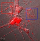
The medium spiny neurons are medium sized neurons with large and extensive dendritic trees. Each branch of the these dendritic trees is packed with numerous small spines which receive synaptic inputs from neurons outside the striatum. The corpus striatum - consisting of nucleus caudatusCaudate nucleus
The caudate nucleus is a nucleus located within the basal ganglia of the brains of many animal species. The caudate nucleus is an important part of the brain's learning and memory system.-Anatomy:...
, putamen, and nucleus accumbens - is the main input station of the basal ganglia. Medium spiny neurons in this structure receive cortical, thalamic
Thalamus
The thalamus is a midline paired symmetrical structure within the brains of vertebrates, including humans. It is situated between the cerebral cortex and midbrain, both in terms of location and neurological connections...
and brain-stem
Brain stem
In vertebrate anatomy the brainstem is the posterior part of the brain, adjoining and structurally continuous with the spinal cord. The brain stem provides the main motor and sensory innervation to the face and neck via the cranial nerves...
inputs. In fact, the whole human neocortex except the primary visual and primary auditory cortex
Primary auditory cortex
The primary auditory cortex is the region of the brain that is responsible for the processing of auditory information. Corresponding roughly with Brodmann areas 41 and 42, it is located on the temporal lobe, and performs the basics of hearing—pitch and volume...
project to the striatum.
Within the striatum, there are at least two different types of medium spiny neurons. These types were first distinguished because of the different neuropeptides they contain. About half the spiny cells express Substance P, dynorphin and dopamine D1 receptors and project to the internal globus pallidus and substantia nigra pars reticulata (the direct pathways) whereas the other half express enkephalin and the dopamine D2 receptor and project to the external globus pallidus (the "indirect pathway"). With different types of immunocytochemical or histochemical staining one can identify small clusters (150-300 µm in diameter) of medium spiny neurons (called "patches" or "striosomes" - making up about 15% of the volume of the neostriatum) embedded in a surrounding "matrix" (making up about 85% of the volume of the striatum).
Function
The medium spiny neurons are GABAergicGamma-aminobutyric acid
γ-Aminobutyric acid is the chief inhibitory neurotransmitter in the mammalian central nervous system. It plays a role in regulating neuronal excitability throughout the nervous system...
neurons and hence have an inhibitory influence on the neurons they project to. Within the basal ganglia, there are several complex circuits of neuronal loops all of which include the medium spiny neurons (for further information see basal ganglia
Basal ganglia
The basal ganglia are a group of nuclei of varied origin in the brains of vertebrates that act as a cohesive functional unit. They are situated at the base of the forebrain and are strongly connected with the cerebral cortex, thalamus and other brain areas...
). They send axons to the internal and external segment of the globus pallidus
Globus pallidus
The globus pallidus also known as paleostriatum, is a sub-cortical structure of the brain. Topographically, it is part of the telencephalon, but retains close functional ties with the subthalamus - both of which are part of the extrapyramidal motor system...
as well as the substantia nigra
Substantia nigra
The substantia nigra is a brain structure located in the mesencephalon that plays an important role in reward, addiction, and movement. Substantia nigra is Latin for "black substance", as parts of the substantia nigra appear darker than neighboring areas due to high levels of melanin in...
pars reticulata.
The cortical, thalamic, and brain-stem inputs that arrive at the medium spiny neurons show a vast divergence in that each incoming axon forms contacts with many spiny neurons and each spiny neuron receives a vast amount of input from different incoming axons. Since these inputs are glutamatergic they exhibit an excitatory influence on the inhibitory medium spiny neurons.
There are also a large number of interneurons originating in different areas which regulate the excitability of the medium spiny neurons. The synaptic connections between the spiny neurons and the interneurons are typically close to the spiny neurons' cell soma, or body. Recall that excitatory postsynaptic potentials caused by glutamatergic inputs at the dendrites of the spiny neurons only cause an action potential
Action potential
In physiology, an action potential is a short-lasting event in which the electrical membrane potential of a cell rapidly rises and falls, following a consistent trajectory. Action potentials occur in several types of animal cells, called excitable cells, which include neurons, muscle cells, and...
when the depolarization wave is strong enough upon entering the cell soma. Since the interneurons' influence is located so closely to this critical gate between the dendrites and the soma, they can readily regulate the generation of an action potential. As a result, the excitatory input coming from cortical etc. neurons has to be very strong, or caused by many simultaneously arriving excitations.
In consequence the medium spiny neurons are usually quiet and do not exhibit any spontaneous activity unless sufficiently activated.
Direct pathway within the basal ganglia
The direct pathway within the basal ganglia makes excitatory inputs coming from e.g. the cortex cause a net excitation of upper motor neuronUpper motor neuron
Upper motor neurons are motor neurons that originate in the motor region of the cerebral cortex or the brain stem and carry motor information down to the final common pathway, that is, any motor neurons that are not directly responsible for stimulating the target muscle...
s in the motor areas of the cortex. In the direct pathway, the medium spiny neurons project to the internal division of the globus pallidus which in turn sends axons to the substantia nigra pars reticulata (SNpr) and the ventroanterior and ventrolateral thalamus (VTh). The SNpr projects to the deep layer of the superior colliculus
Superior colliculus
The optic tectum or simply tectum is a paired structure that forms a major component of the vertebrate midbrain. In mammals this structure is more commonly called the superior colliculus , but, even in mammals, the adjective tectal is commonly used. The tectum is a layered structure, with a...
thus controlling fast eye movements (saccades). The VTh projects to upper motor neurons in the primary motor cortex
Primary motor cortex
The primary motor cortex is a brain region that in humans is located in the posterior portion of the frontal lobe. Itworks in association with pre-motor areas to plan and execute movements. M1 contains large neurons known as Betz cells, which send long axons down the spinal cord to synapse onto...
(precentral gyrus).
Neurons in the globus pallidus are also inhibitory, thus inhibiting the excitatory neurons in the SNpr and VTh. But in contrast to the medium spiny neurons, globus pallidus neurons are tonically active when not activated. Thus in the absence of cortical stimulation, SNpr and VTh neurons are tonically inhibited thus preventing involuntary spontaneous movements.
Once the medium spiny neurons receive sufficient excitatory cortical input, they are excited and fire a burst of inhibitory action potentials to globus pallidus neurons. These tonically active neurons are then inhibited, causing their inhibitory influence on SNpr and VTh to decline. Thus SNpr and VTh neurons are disinhibited resulting in net excitement causing them to activate upper motor neurons commanding a movement. Cortical activation of the basal ganglia thus eventually results in excitement (disinhibition
Disinhibition
Disinhibition is a term in psychology used to describe a lack of restraint manifested in several ways, including disregard for social conventions, impulsivity, and poor risk assessment. Disinhibition affects motor, instinctual, emotional, cognitive and perceptual aspects with signs and symptoms...
) of motor neuron
Motor neuron
In vertebrates, the term motor neuron classically applies to neurons located in the central nervous system that project their axons outside the CNS and directly or indirectly control muscles...
s causing movement to take place.
Indirect pathway
In the indirect pathway, excitatory e.g. cortical input to the basal ganglia results in net inhibition of upper motor neurons. In this pathway the medium spiny neurons in the striatum project to the external segment of the globus pallidus. These neurons in turn project to the internal segment of the globus pallidus and to the subthalamic nuclei which form a closed loopPID controller
A proportional–integral–derivative controller is a generic control loop feedback mechanism widely used in industrial control systems – a PID is the most commonly used feedback controller. A PID controller calculates an "error" value as the difference between a measured process variable and a...
by projecting back to the internal globus pallidus.
Cortical excitement of medium spiny neurons causes them to inhibit external globus pallidus neurons. These tonically inhibiting neurons thus decrease their inhibitory influence on the internal globus pallidus and the subthalamic nuclei.
Let's first look at the internal globus pallidus neurons which are also tonically inhibiting VTh and SNpr neurons. Since the inhibitory influence from the external globus pallidus is now reduced, these neurons show stronger activity thus increasing their inhibition of SNpr and VTh neurons.
The projections of the external globus pallidus to the subthalamic nuclei causes these neurons to increase their firing rate, since the globus pallidus neurons are inhibited by medium spiny neurons. The subthalamic nuclei have excitatory projections to the internal globus pallidus thus causing the internal globus pallidus neurons to increase their inhibititory influence on SNpr and VTh.
Eventually excitatory inputs from the cortex results in net inhibition of upper motor neurons thus preventing them from initiating a movement.
External links
- NIF Search - Medium Spiny Neuron via the Neuroscience Information FrameworkNeuroscience Information FrameworkThe Neuroscience Information Framework is a repository of global neuroscience web resources, including experimental, clinical, and translational neuroscience databases, knowledge bases, atlases, and genetic/genomic resources.-Description:...

