Radiography
Encyclopedia

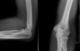
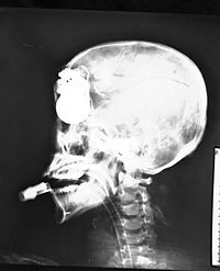
A heterogeneous beam of X-rays is produced by an X-ray generator and is projected toward an object. According to the density and composition of the different areas of the object a proportion of X-rays are absorbed by the object. The X-rays that pass through are then captured behind the object by a detector (film sensitive to X-rays or a digital detector) which gives a 2D representation of all the structures superimposed on each other. In tomography
Tomography
Tomography refers to imaging by sections or sectioning, through the use of any kind of penetrating wave. A device used in tomography is called a tomograph, while the image produced is a tomogram. The method is used in radiology, archaeology, biology, geophysics, oceanography, materials science,...
, the X-ray source and detector move to blur out structures not in the focal plane. Computed tomography
Computed tomography
X-ray computed tomography or Computer tomography , is a medical imaging method employing tomography created by computer processing...
(CT scanning) is different to plain film tomography in that computer assisted reconstruction is used to generate a 3D representation of the scanned object/patient.
Medical and industrial radiography
Radiography is used for both medical and industrialIndustry
Industry refers to the production of an economic good or service within an economy.-Industrial sectors:There are four key industrial economic sectors: the primary sector, largely raw material extraction industries such as mining and farming; the secondary sector, involving refining, construction,...
applications (see medical radiography
Medical radiography
Radiography is the use of ionizing electromagnetic radiation such as X-rays to view objects. Although not technically radiographic techniques, imaging modalities such as PET and MRI are sometimes grouped in radiography because the radiology department of hospitals handle all forms of imaging...
and industrial radiography
Industrial radiography
Industrial Radiography is the use of ionizing radiation to view objects in a way that cannot be seen otherwise. It is not to be confused with the use of ionizing radiation to change or modify objects; radiography's purpose is strictly viewing. Industrial radiography has grown out of engineering,...
). If the object being examined is living, whether human
Human
Humans are the only living species in the Homo genus...
or animal
Animal
Animals are a major group of multicellular, eukaryotic organisms of the kingdom Animalia or Metazoa. Their body plan eventually becomes fixed as they develop, although some undergo a process of metamorphosis later on in their life. Most animals are motile, meaning they can move spontaneously and...
, it is regarded as medical; all other radiography is regarded as industrial radiographic work.
History of radiography

X-ray
X-radiation is a form of electromagnetic radiation. X-rays have a wavelength in the range of 0.01 to 10 nanometers, corresponding to frequencies in the range 30 petahertz to 30 exahertz and energies in the range 120 eV to 120 keV. They are shorter in wavelength than UV rays and longer than gamma...
s, also referred to as Röntgen rays after Wilhelm Conrad Röntgen
Wilhelm Conrad Röntgen
Wilhelm Conrad Röntgen was a German physicist, who, on 8 November 1895, produced and detected electromagnetic radiation in a wavelength range today known as X-rays or Röntgen rays, an achievement that earned him the first Nobel Prize in Physics in 1901....
who first described their properties in rigorous detail. These previously unknown rays (hence the X) were found to be a type of electromagnetic radiation
Electromagnetic radiation
Electromagnetic radiation is a form of energy that exhibits wave-like behavior as it travels through space...
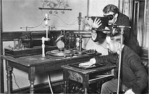
. It wasn't long before X-rays were used in various applications, from helping to fit shoes to the medical uses of today. The first radiograph used to assist in surgery was taken a year after its invention in Birmingham
Birmingham
Birmingham is a city and metropolitan borough in the West Midlands of England. It is the most populous British city outside the capital London, with a population of 1,036,900 , and lies at the heart of the West Midlands conurbation, the second most populous urban area in the United Kingdom with a...
by the British pioneer of medical X-Rays, Major John Hall-Edwards
John Hall-Edwards
John Francis Hall-Edwards was a pioneer in the medical use of X-rays in the United Kingdom. He took the first radiograph to direct a surgical operation, on 14 February 1896. Hall-Edwards' interest in X-rays cost him his left arm, which had to be amputated in 1908 as a consequence of X-ray...
. X-rays were put to diagnostic use very early, before the dangers of ionizing radiation were discovered. Indeed, Marie Curie
Marie Curie
Marie Skłodowska-Curie was a physicist and chemist famous for her pioneering research on radioactivity. She was the first person honored with two Nobel Prizes—in physics and chemistry...
pushed for radiography to be used to treat wounded soldiers in World War I. Initially, many kinds of staff conducted radiography in hospitals, including physicists, photographers, doctors, nurses, and engineers. The medical specialty of radiology grew up over many years around the new technology. When new diagnostic tests were developed, it was natural for the radiographers
Radiologic technologist
A radiologic technologist, also known as medical radiation technologist and as radiographer, performs imaging of the human body for diagnosis or treating medical problems...
to be trained in and to adopt this new technology. Radiographers now often do fluoroscopy
Fluoroscopy
Fluoroscopy is an imaging technique commonly used by physicians to obtain real-time moving images of the internal structures of a patient through the use of a fluoroscope. In its simplest form, a fluoroscope consists of an X-ray source and fluorescent screen between which a patient is placed...
, computed tomography
Computed tomography
X-ray computed tomography or Computer tomography , is a medical imaging method employing tomography created by computer processing...
, mammography
Mammography
Mammography is the process of using low-energy-X-rays to examine the human breast and is used as a diagnostic and a screening tool....
, ultrasound
Ultrasound
Ultrasound is cyclic sound pressure with a frequency greater than the upper limit of human hearing. Ultrasound is thus not separated from "normal" sound based on differences in physical properties, only the fact that humans cannot hear it. Although this limit varies from person to person, it is...
, nuclear medicine
Nuclear medicine
In nuclear medicine procedures, elemental radionuclides are combined with other elements to form chemical compounds, or else combined with existing pharmaceutical compounds, to form radiopharmaceuticals. These radiopharmaceuticals, once administered to the patient, can localize to specific organs...
and magnetic resonance imaging
Magnetic resonance imaging
Magnetic resonance imaging , nuclear magnetic resonance imaging , or magnetic resonance tomography is a medical imaging technique used in radiology to visualize detailed internal structures...
as well. Although a nonspecialist dictionary might define radiography quite narrowly as "taking X-ray images", this has long been only part of the work of "X-ray departments", radiographers, and radiologists. Initially, radiographs were known as roentgenograms.
Equipment

Sources
A number of sources of X-rayX-ray
X-radiation is a form of electromagnetic radiation. X-rays have a wavelength in the range of 0.01 to 10 nanometers, corresponding to frequencies in the range 30 petahertz to 30 exahertz and energies in the range 120 eV to 120 keV. They are shorter in wavelength than UV rays and longer than gamma...
photon
Photon
In physics, a photon is an elementary particle, the quantum of the electromagnetic interaction and the basic unit of light and all other forms of electromagnetic radiation. It is also the force carrier for the electromagnetic force...
s have been used; these include X-ray generators, betatron
Betatron
A betatron is a cyclotron developed by Donald Kerst at the University of Illinois in 1940 to accelerate electrons, but the concepts ultimately originate from Rolf Widerøe and previous development occurred in Germany through Max Steenbeck in the 1930s. The betatron is essentially a transformer with...
s, and linear accelerators
Linear particle accelerator
A linear particle accelerator is a type of particle accelerator that greatly increases the velocity of charged subatomic particles or ions by subjecting the charged particles to a series of oscillating electric potentials along a linear beamline; this method of particle acceleration was invented...
(linacs). For gamma ray
Gamma ray
Gamma radiation, also known as gamma rays or hyphenated as gamma-rays and denoted as γ, is electromagnetic radiation of high frequency . Gamma rays are usually naturally produced on Earth by decay of high energy states in atomic nuclei...
s, radioactive sources such as 192Ir, 60Co
Cobalt-60
Cobalt-60, , is a synthetic radioactive isotope of cobalt. Due to its half-life of 5.27 years, is not found in nature. It is produced artificially by neutron activation of . decays by beta decay to the stable isotope nickel-60...
or 137Cs
Caesium-137
Caesium-137 is a radioactive isotope of caesium which is formed as a fission product by nuclear fission.It has a half-life of about 30.17 years, and decays by beta emission to a metastable nuclear isomer of barium-137: barium-137m . Caesium-137 is a radioactive isotope of caesium which is formed...
are used.
Detectors
A range of detectors including photographic filmPhotographic film
Photographic film is a sheet of plastic coated with an emulsion containing light-sensitive silver halide salts with variable crystal sizes that determine the sensitivity, contrast and resolution of the film...
, scintillator
Scintillator
A scintillator is a special material, which exhibits scintillation—the property of luminescence when excited by ionizing radiation. Luminescent materials, when struck by an incoming particle, absorb its energy and scintillate, i.e., reemit the absorbed energy in the form of light...
and semiconductor
Semiconductor
A semiconductor is a material with electrical conductivity due to electron flow intermediate in magnitude between that of a conductor and an insulator. This means a conductivity roughly in the range of 103 to 10−8 siemens per centimeter...
diode
Diode
In electronics, a diode is a type of two-terminal electronic component with a nonlinear current–voltage characteristic. A semiconductor diode, the most common type today, is a crystalline piece of semiconductor material connected to two electrical terminals...
arrays have been used to collect images.
Theory of X-ray attenuation
X-ray photons used for medical purposes are formed by an event involving an electron, while gamma ray photons are formed from an interaction with the nucleus of an atom. In general, medical radiography is done using X-rays formed in an X-ray tubeX-ray tube
An X-ray tube is a vacuum tube that produces X-rays. They are used in X-ray machines. X-rays are part of the electromagnetic spectrum, an ionizing radiation with wavelengths shorter than ultraviolet light...
. Nuclear medicine typically involves gamma rays.
The types of electromagnetic radiation
Electromagnetic radiation
Electromagnetic radiation is a form of energy that exhibits wave-like behavior as it travels through space...
of most interest to radiography are X-ray and gamma radiation. This radiation is much more energetic
Energy
In physics, energy is an indirectly observed quantity. It is often understood as the ability a physical system has to do work on other physical systems...
than the more familiar types such as radio waves
Radio waves
Radio waves are a type of electromagnetic radiation with wavelengths in the electromagnetic spectrum longer than infrared light. Radio waves have frequencies from 300 GHz to as low as 3 kHz, and corresponding wavelengths from 1 millimeter to 100 kilometers. Like all other electromagnetic waves,...
and visible light. It is this relatively high energy which makes gamma rays useful in radiography but potentially hazardous to living organisms.

Radium
Radium is a chemical element with atomic number 88, represented by the symbol Ra. Radium is an almost pure-white alkaline earth metal, but it readily oxidizes on exposure to air, becoming black in color. All isotopes of radium are highly radioactive, with the most stable isotope being radium-226,...
and radon
Radon
Radon is a chemical element with symbol Rn and atomic number 86. It is a radioactive, colorless, odorless, tasteless noble gas, occurring naturally as the decay product of uranium or thorium. Its most stable isotope, 222Rn, has a half-life of 3.8 days...
, and artificially produced radioactive isotopes of elements, such as cobalt-60 and iridium-192. Electromagnetic radiation consists of oscillating
Oscillation
Oscillation is the repetitive variation, typically in time, of some measure about a central value or between two or more different states. Familiar examples include a swinging pendulum and AC power. The term vibration is sometimes used more narrowly to mean a mechanical oscillation but sometimes...
electric
Electric field
In physics, an electric field surrounds electrically charged particles and time-varying magnetic fields. The electric field depicts the force exerted on other electrically charged objects by the electrically charged particle the field is surrounding...
and magnetic
Magnetic field
A magnetic field is a mathematical description of the magnetic influence of electric currents and magnetic materials. The magnetic field at any given point is specified by both a direction and a magnitude ; as such it is a vector field.Technically, a magnetic field is a pseudo vector;...
fields, but is generally depicted as a single sinusoidal wave. While in the past radium
Radium
Radium is a chemical element with atomic number 88, represented by the symbol Ra. Radium is an almost pure-white alkaline earth metal, but it readily oxidizes on exposure to air, becoming black in color. All isotopes of radium are highly radioactive, with the most stable isotope being radium-226,...
and radon
Radon
Radon is a chemical element with symbol Rn and atomic number 86. It is a radioactive, colorless, odorless, tasteless noble gas, occurring naturally as the decay product of uranium or thorium. Its most stable isotope, 222Rn, has a half-life of 3.8 days...
have both been used for radiography, they have fallen out of use as they are radiotoxic alpha radiation emitters which are expensive; iridium-192 and cobalt-60 are far better photon sources. For further details see commonly used gamma emitting isotopes
Commonly used gamma emitting isotopes
Radionuclides which emit gamma radiation are valuable in a range of different industrial, scientific and medical technologies. This article lists some common gamma-emitting radionuclides of technological importance, and their properties.- Fission products :...
.
Gamma rays are indirectly ionizing radiation
Ionizing radiation
Ionizing radiation is radiation composed of particles that individually have sufficient energy to remove an electron from an atom or molecule. This ionization produces free radicals, which are atoms or molecules containing unpaired electrons...
. A gamma ray passes through matter
Matter
Matter is a general term for the substance of which all physical objects consist. Typically, matter includes atoms and other particles which have mass. A common way of defining matter is as anything that has mass and occupies volume...
until it undergoes an interaction with an atom
Atom
The atom is a basic unit of matter that consists of a dense central nucleus surrounded by a cloud of negatively charged electrons. The atomic nucleus contains a mix of positively charged protons and electrically neutral neutrons...
ic particle, usually an electron
Electron
The electron is a subatomic particle with a negative elementary electric charge. It has no known components or substructure; in other words, it is generally thought to be an elementary particle. An electron has a mass that is approximately 1/1836 that of the proton...
. During this interaction, energy is transferred from the gamma ray to the electron, which is a directly ionizing particle. As a result of this energy transfer, the electron is liberated from the atom and proceeds to ionize matter by colliding with other electrons along its path. Other times, the passing gamma ray interferes with the orbit of the electron, and slows it, releasing energy but not becoming dislodged. The atom is not ionised, and the gamma ray continues on, although at a lower energy. This energy released is usually heat or another, weaker photon, and causes biological harm as a radiation burn. The chain reaction caused by the initial dose of radiation can continue after exposure, much like a sunburn
Sunburn
A sunburn is a burn to living tissue, such as skin, which is produced by overexposure to ultraviolet radiation, commonly from the sun's rays. Usual mild symptoms in humans and other animals include red or reddish skin that is hot to the touch, general fatigue, and mild dizziness. An excess of UV...
continues to damage skin even after one is out of direct sunlight.
For the range of energies commonly used in radiography, the interaction between gamma rays and electrons occurs in two ways. One effect takes place where all the gamma ray's energy is transmitted to an entire atom. The gamma ray no longer exists and an electron emerges from the atom with kinetic
Kinetic energy
The kinetic energy of an object is the energy which it possesses due to its motion.It is defined as the work needed to accelerate a body of a given mass from rest to its stated velocity. Having gained this energy during its acceleration, the body maintains this kinetic energy unless its speed changes...
(motion in relation to force) energy almost equal to the gamma energy. This effect is predominant at low gamma energies and is known as the photoelectric effect
Photoelectric effect
In the photoelectric effect, electrons are emitted from matter as a consequence of their absorption of energy from electromagnetic radiation of very short wavelength, such as visible or ultraviolet light. Electrons emitted in this manner may be referred to as photoelectrons...
. The other major effect occurs when a gamma ray interacts with an atomic electron, freeing it from the atom and imparting to it only a fraction of the gamma ray's kinetic energy. A secondary gamma ray with less energy (hence lower frequency) also emerges from the interaction. This effect predominates at higher gamma energies and is known as the Compton effect.
In both of these effects the emergent electrons lose their kinetic energy by ionizing surrounding atoms. The density of ion
Ion
An ion is an atom or molecule in which the total number of electrons is not equal to the total number of protons, giving it a net positive or negative electrical charge. The name was given by physicist Michael Faraday for the substances that allow a current to pass between electrodes in a...
s so generated is a measure of the energy delivered to the material by the gamma rays.
The most common means of measuring the variations in a beam of radiation is by observing its effect on a photographic film. This effect is the same as that of light, and the more intense the radiation is, the more it darkens, or exposes
Exposure (photography)
In photography, exposure is the total amount of light allowed to fall on the photographic medium during the process of taking a photograph. Exposure is measured in lux seconds, and can be computed from exposure value and scene luminance over a specified area.In photographic jargon, an exposure...
, the film. Other methods are in use, such as the ionizing effect measured electronically, its ability to discharge an electrostatically charged plate or to cause certain chemicals to fluoresce as in fluoroscopy
Fluoroscopy
Fluoroscopy is an imaging technique commonly used by physicians to obtain real-time moving images of the internal structures of a patient through the use of a fluoroscope. In its simplest form, a fluoroscope consists of an X-ray source and fluorescent screen between which a patient is placed...
.
Obsolete terminology
The term skiagrapher was used until about 1918 to mean radiographer. It was derived from Ancient GreekAncient Greek
Ancient Greek is the stage of the Greek language in the periods spanning the times c. 9th–6th centuries BC, , c. 5th–4th centuries BC , and the c. 3rd century BC – 6th century AD of ancient Greece and the ancient world; being predated in the 2nd millennium BC by Mycenaean Greek...
words for 'shadow' and 'writer'.
See also
- Computer-aided diagnosisComputer-aided diagnosisComputer-aided detection and computer-aided diagnosis are procedures in medicine that assist doctors in the interpretation of medical images. Imaging techniques in X-ray, MRI, and Ultrasound diagnostics yield a great deal of information, which the radiologist has to analyze and evaluate...
- GenitographyGenitographyGenitography is the radiography of the urogenital sinus and internal duct structures after injection of a contrast medium through the opening of the sinus.-References:* Dorland's Illustrated Medical Dictionary...
- RadiationRadiationIn physics, radiation is a process in which energetic particles or energetic waves travel through a medium or space. There are two distinct types of radiation; ionizing and non-ionizing...
- Radiation contamination
- List of civilian radiation accidents
- Radiographer
- Projectional radiographyProjectional radiographyProjectional radiography or plain film radiography is the practice of producing two-dimensional images using x-ray radiation. Radiographic exams are typically performed by Radiologic Technologists, highly trained medical professionals who specialize in the usage of radiographic equipment, patient...
- Shadowgraphs
- Background radiationBackground radiationBackground radiation is the ionizing radiation constantly present in the natural environment of the Earth, which is emitted by natural and artificial sources.-Overview:Both Natural and human-made background radiation varies by location....
External links
- MedPix Medical Image Database
- NIST's XAAMDI: X-Ray Attenuation and Absorption for Materials of Dosimetric Interest Database
- NIST's XCOM: Photon Cross Sections Database
- NIST's FAST: Attenuation and Scattering Tables
- Australian Institute of Radiography
- American College of Radiology
- UN information on the security of industrial sources
- European Society of Radiology
- What is Radiology?
- New York State Society of Radiologic Technologists

