Angle-resolved low-coherence interferometry
Encyclopedia
For the electrical device, see ALCI
Angle-resolved low-coherence interferometry (a/LCI) is an emerging biomedical imaging
technology which uses the properties of scattered light
to measure the average size of cell structures, including cell nuclei
. The technology shows promise as a clinical tool for in situ detection of dysplatic
, or precancerous
tissue.
to solve the inverse problem
of determining scatterer geometry based on far field diffraction patterns. Similar to optical coherence domain reflectometry (OCDR) and optical coherence tomography
(OCT), a/LCI uses a broadband light source in an interferometry
scheme in order to achieve optical sectioning with a depth resolution set by the coherence length
of the source. Angle-resolved scattering measurements capture light
as a function of the scattering angle, and invert the angles to deduce the average size of the scattering objects via a computational light scattering model
such as Mie theory
, which predicts angles based on the size of the scattering sphere
. Combining these techniques allows construction of a system that can measure average scatter size at various depths within a tissue sample
.
At present the most significant medical application of the technology is determining the state of tissue health based on measurements of average cell nuclei size. It has been found that as tissue changes from normal to cancerous, the average cell nuclei size increases. Several recent studies have shown that via cell nuclei measurements, a/LCI can detect the presence of low- and high-grade dysplasia with 91% sensitivity and distinguish between normal and dysplastic with 97% specificity.
as well as the diagnosis of dysplasia
. Variations in scattering distributions as a function of angle
or wavelength
have been used to deduce information regarding the size of cells and subcellular objects such as nuclei and organelle
s. These size measurements can then be used diagnostically to detect tissue changes—including neoplastic
changes (those leading to cancer).
Light scattering spectroscopy has been used to detect dysplasia in the colon
, bladder
, cervix
, and esophagus
of human patients. Light scattering has also been used to detect Barrett’s esophagus, a metaplastic condition with a high probability of leading to dysplasia.
However, in contrast with a/LCI, these techniques all rely on total intensity based measurements, which lack the ability to provide results as a function of depth in the tissue.
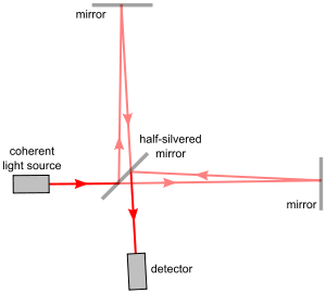
 The first implementation of a/LCI used a Michelson interferometer
, the same model used in the famous Michelson-Morley experiment
. The Michelson interferometer splits one beam of light into two paths, one reference path and one sampling path, and recombines them again to produce a waveform resulting from interference. The difference between the reference beam and the sampling beam thus reveal the properties of the sample in the way it scatters light.
The early a/LCI device used a movable mirror and lens in the reference arm so that researchers could replicate different angles and depths in the reference beam as they occurred in the collected backscattered light. This allowed isolation of the backscattered light at varying depths of reflection in the sample.
In order to transform the data into measurements of cell structure, angular scattering distributions are then compared to the predictions of Mie theory
—which calculates the size of spheres relative to their light scattering patterns.
The a/LCI technique was first validated in studies of polystyrene microspheres, the sizes of which were known and relatively homogenous. A later study expanded the signal processing method to compensate for the nonspherical and inhomogeneous nature of cell nuclei.
This early system required up to 40 minutes to acquire the data for a 1 mm² point in a sample, but proved the feasibility of the idea.
 Like OCT, the early implementations of a/LCI relied on physically changing the optical path length
(OPL) to control the depth in the sample from which data are acquired. However, it has been demonstrated that it is possible to use a Fourier domain implementation to yield depth resolution in a single data acquisition. A broadband
light source is used to produce a spectrum of wavelengths at once, and the backscattered light is collected by a coherent
optical fiber
in the return path to capture different scattering angles simultaneously. Intensity is then measured via a spectrometer
: a single frame from the spectrometer contains scattering intensity as a function of wavelength
and angle. Finally the data is Fourier transform
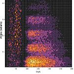
ed on a line-by-line basis to generate scattering intensity as a function of OPL and angle. In the resulting image, the x axis represents the OPL and the y axis the angle of reflection, thus yielding a 2D map of reflection intensities.
Using this method, the acquisition speed is limited only by the integration time of the spectrometer and can be as short at 20 ms. The same data that initially required tens of minutes to acquire can be acquired ~105 times faster.
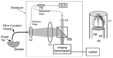
 The Fourier-domain version of the a/LCI system uses a superluminescent diode
(SLD) with a fiber-coupled output as the light source. A fiber splitter separates the signal path at 90% intensity and the reference path at 10%.
The light from the SLD passes through an optical isolator
and subsequently a polarization controller
. It has been shown that control of light polarization is important for maximizing optical signal and comparing angular scattering with the Mie scattering model. A polarization-maintaining fiber is used to carry the illumination light to the sample. A second polarization controller is similarly used to control the polarization of the light passing through the reference path.
The output of the fiber on the right is collimated
using lens L1 and illuminates the tissue. But because the delivery fiber is offset from the optical axis of the lens, the beam is delivered to the sample at an oblique angle. Backscattered light is then collimated by the same lens and collected by the fiber bundle. The fibers are one focal length from the lens, and the sample is one focal length on the other side. This configuration captures light from the maximum range of angles and minimizes light noise due to specular reflections.
At the distal end of the fiber bundle, light from each fiber is imaged onto the spectrometer. Light from the sample and reference arms are mixed by a beamsplitting
cube (BS), and are incident on the entrance slit of an imaging spectrometer. Data from the imaging spectrometer are transferred to a computer via universal serial bus
interface for signal processing and display of results. The computer also provides control of the imaging spectrometer.
ball lens in the probe tip, which reduces reflections that otherwise limit the depth range of the system.
The portable system uses a 2 ft by 2 ft optical breadboard
as the base, with the source, fiber optic components, lens, beamsplitter, and imaging spectrometer mounted to the breadboard. An aluminum cover protects the optics. A fiber probe with a handheld probe enables easy access to tissue samples for testing. On the left side sits a white sample platform, where tissue is placed for testing. The handheld probe is used by the operator to select specific sites on the tissue from which a/LCI readings are acquired.
Residual-current device
A Residual Current Device is a generic term covering both RCCBs and RCBOs.A Residual-Current Circuit Breaker is an electrical wiring device that disconnects a circuit whenever it detects that the electric current is not balanced between the energized conductor and the return neutral conductor...
Angle-resolved low-coherence interferometry (a/LCI) is an emerging biomedical imaging
Medical imaging
Medical imaging is the technique and process used to create images of the human body for clinical purposes or medical science...
technology which uses the properties of scattered light
Scattering
Scattering is a general physical process where some forms of radiation, such as light, sound, or moving particles, are forced to deviate from a straight trajectory by one or more localized non-uniformities in the medium through which they pass. In conventional use, this also includes deviation of...
to measure the average size of cell structures, including cell nuclei
Cell nucleus
In cell biology, the nucleus is a membrane-enclosed organelle found in eukaryotic cells. It contains most of the cell's genetic material, organized as multiple long linear DNA molecules in complex with a large variety of proteins, such as histones, to form chromosomes. The genes within these...
. The technology shows promise as a clinical tool for in situ detection of dysplatic
Dysplasia
Dysplasia , is a term used in pathology to refer to an abnormality of development. This generally consists of an expansion of immature cells, with a corresponding decrease in the number and location of mature cells. Dysplasia is often indicative of an early neoplastic process...
, or precancerous
Carcinoma in situ
Carcinoma in situ is an early form of cancer that is defined by the absence of invasion of tumor cells into the surrounding tissue, usually before penetration through the basement membrane. In other words, the neoplastic cells proliferate in their normal habitat, hence the name "in situ"...
tissue.
Introduction
A/LCI combines low-coherence interferometry with angle-resolved scatteringScattering
Scattering is a general physical process where some forms of radiation, such as light, sound, or moving particles, are forced to deviate from a straight trajectory by one or more localized non-uniformities in the medium through which they pass. In conventional use, this also includes deviation of...
to solve the inverse problem
Inverse problem
An inverse problem is a general framework that is used to convert observed measurements into information about a physical object or system that we are interested in...
of determining scatterer geometry based on far field diffraction patterns. Similar to optical coherence domain reflectometry (OCDR) and optical coherence tomography
Optical coherence tomography
Optical coherence tomography is an optical signal acquisition and processing method. It captures micrometer-resolution, three-dimensional images from within optical scattering media . Optical coherence tomography is an interferometric technique, typically employing near-infrared light...
(OCT), a/LCI uses a broadband light source in an interferometry
Interferometry
Interferometry refers to a family of techniques in which electromagnetic waves are superimposed in order to extract information about the waves. An instrument used to interfere waves is called an interferometer. Interferometry is an important investigative technique in the fields of astronomy,...
scheme in order to achieve optical sectioning with a depth resolution set by the coherence length
Coherence length
In physics, coherence length is the propagation distance from a coherent source to a point where an electromagnetic wave maintains a specified degree of coherence. The significance is that interference will be strong within a coherence length of the source, but not beyond it...
of the source. Angle-resolved scattering measurements capture light
Light
Light or visible light is electromagnetic radiation that is visible to the human eye, and is responsible for the sense of sight. Visible light has wavelength in a range from about 380 nanometres to about 740 nm, with a frequency range of about 405 THz to 790 THz...
as a function of the scattering angle, and invert the angles to deduce the average size of the scattering objects via a computational light scattering model
Light scattering by particles
Light scattering by particles is the process by which small particles such as ice crystals, dust, planetary dust, and blood cells cause observable phenomena such as rainbows, the color of the sky, and halos....
such as Mie theory
Mie theory
The Mie solution to Maxwell's equations describes the scattering of electromagnetic radiation by a sphere...
, which predicts angles based on the size of the scattering sphere
Sphere
A sphere is a perfectly round geometrical object in three-dimensional space, such as the shape of a round ball. Like a circle in two dimensions, a perfect sphere is completely symmetrical around its center, with all points on the surface lying the same distance r from the center point...
. Combining these techniques allows construction of a system that can measure average scatter size at various depths within a tissue sample
Biopsy
A biopsy is a medical test involving sampling of cells or tissues for examination. It is the medical removal of tissue from a living subject to determine the presence or extent of a disease. The tissue is generally examined under a microscope by a pathologist, and can also be analyzed chemically...
.
At present the most significant medical application of the technology is determining the state of tissue health based on measurements of average cell nuclei size. It has been found that as tissue changes from normal to cancerous, the average cell nuclei size increases. Several recent studies have shown that via cell nuclei measurements, a/LCI can detect the presence of low- and high-grade dysplasia with 91% sensitivity and distinguish between normal and dysplastic with 97% specificity.
History
Since 2000, light scattering systems have been used for biomedical applications such as the study of cellular morphologyMorphology (biology)
In biology, morphology is a branch of bioscience dealing with the study of the form and structure of organisms and their specific structural features....
as well as the diagnosis of dysplasia
Dysplasia
Dysplasia , is a term used in pathology to refer to an abnormality of development. This generally consists of an expansion of immature cells, with a corresponding decrease in the number and location of mature cells. Dysplasia is often indicative of an early neoplastic process...
. Variations in scattering distributions as a function of angle
Angle
In geometry, an angle is the figure formed by two rays sharing a common endpoint, called the vertex of the angle.Angles are usually presumed to be in a Euclidean plane with the circle taken for standard with regard to direction. In fact, an angle is frequently viewed as a measure of an circular arc...
or wavelength
Wavelength
In physics, the wavelength of a sinusoidal wave is the spatial period of the wave—the distance over which the wave's shape repeats.It is usually determined by considering the distance between consecutive corresponding points of the same phase, such as crests, troughs, or zero crossings, and is a...
have been used to deduce information regarding the size of cells and subcellular objects such as nuclei and organelle
Organelle
In cell biology, an organelle is a specialized subunit within a cell that has a specific function, and is usually separately enclosed within its own lipid bilayer....
s. These size measurements can then be used diagnostically to detect tissue changes—including neoplastic
Neoplasia
Neoplasm is an abnormal mass of tissue as a result of neoplasia. Neoplasia is the abnormal proliferation of cells. The growth of neoplastic cells exceeds and is not coordinated with that of the normal tissues around it. The growth persists in the same excessive manner even after cessation of the...
changes (those leading to cancer).
Light scattering spectroscopy has been used to detect dysplasia in the colon
Colon (anatomy)
The colon is the last part of the digestive system in most vertebrates; it extracts water and salt from solid wastes before they are eliminated from the body, and is the site in which flora-aided fermentation of unabsorbed material occurs. Unlike the small intestine, the colon does not play a...
, bladder
Bladder
Bladder usually refers to an anatomical hollow organBladder may also refer to:-Biology:* Urinary bladder in humans** Urinary bladder ** Bladder control; see Urinary incontinence** Artificial urinary bladder, in humans...
, cervix
Cervix
The cervix is the lower, narrow portion of the uterus where it joins with the top end of the vagina. It is cylindrical or conical in shape and protrudes through the upper anterior vaginal wall...
, and esophagus
Esophagus
The esophagus is an organ in vertebrates which consists of a muscular tube through which food passes from the pharynx to the stomach. During swallowing, food passes from the mouth through the pharynx into the esophagus and travels via peristalsis to the stomach...
of human patients. Light scattering has also been used to detect Barrett’s esophagus, a metaplastic condition with a high probability of leading to dysplasia.
However, in contrast with a/LCI, these techniques all rely on total intensity based measurements, which lack the ability to provide results as a function of depth in the tissue.
Early a/LCI models

Michelson interferometer
The Michelson interferometer is the most common configuration for optical interferometry and was invented by Albert Abraham Michelson. An interference pattern is produced by splitting a beam of light into two paths, bouncing the beams back and recombining them...
, the same model used in the famous Michelson-Morley experiment
Michelson-Morley experiment
The Michelson–Morley experiment was performed in 1887 by Albert Michelson and Edward Morley at what is now Case Western Reserve University in Cleveland, Ohio. Its results are generally considered to be the first strong evidence against the theory of a luminiferous ether and in favor of special...
. The Michelson interferometer splits one beam of light into two paths, one reference path and one sampling path, and recombines them again to produce a waveform resulting from interference. The difference between the reference beam and the sampling beam thus reveal the properties of the sample in the way it scatters light.
The early a/LCI device used a movable mirror and lens in the reference arm so that researchers could replicate different angles and depths in the reference beam as they occurred in the collected backscattered light. This allowed isolation of the backscattered light at varying depths of reflection in the sample.
In order to transform the data into measurements of cell structure, angular scattering distributions are then compared to the predictions of Mie theory
Mie theory
The Mie solution to Maxwell's equations describes the scattering of electromagnetic radiation by a sphere...
—which calculates the size of spheres relative to their light scattering patterns.
The a/LCI technique was first validated in studies of polystyrene microspheres, the sizes of which were known and relatively homogenous. A later study expanded the signal processing method to compensate for the nonspherical and inhomogeneous nature of cell nuclei.
This early system required up to 40 minutes to acquire the data for a 1 mm² point in a sample, but proved the feasibility of the idea.
Fourier-domain implementation

Optical path length
In optics, optical path length or optical distance is the product of the geometric length of the path light follows through the system, and the index of refraction of the medium through which it propagates. A difference in optical path length between two paths is often called the optical path...
(OPL) to control the depth in the sample from which data are acquired. However, it has been demonstrated that it is possible to use a Fourier domain implementation to yield depth resolution in a single data acquisition. A broadband
Broadband
The term broadband refers to a telecommunications signal or device of greater bandwidth, in some sense, than another standard or usual signal or device . Different criteria for "broad" have been applied in different contexts and at different times...
light source is used to produce a spectrum of wavelengths at once, and the backscattered light is collected by a coherent
Coherence (physics)
In physics, coherence is a property of waves that enables stationary interference. More generally, coherence describes all properties of the correlation between physical quantities of a wave....
optical fiber
Optical fiber
An optical fiber is a flexible, transparent fiber made of a pure glass not much wider than a human hair. It functions as a waveguide, or "light pipe", to transmit light between the two ends of the fiber. The field of applied science and engineering concerned with the design and application of...
in the return path to capture different scattering angles simultaneously. Intensity is then measured via a spectrometer
Spectrometer
A spectrometer is an instrument used to measure properties of light over a specific portion of the electromagnetic spectrum, typically used in spectroscopic analysis to identify materials. The variable measured is most often the light's intensity but could also, for instance, be the polarization...
: a single frame from the spectrometer contains scattering intensity as a function of wavelength
Wavelength
In physics, the wavelength of a sinusoidal wave is the spatial period of the wave—the distance over which the wave's shape repeats.It is usually determined by considering the distance between consecutive corresponding points of the same phase, such as crests, troughs, or zero crossings, and is a...
and angle. Finally the data is Fourier transform
Fourier transform
In mathematics, Fourier analysis is a subject area which grew from the study of Fourier series. The subject began with the study of the way general functions may be represented by sums of simpler trigonometric functions...
ed on a line-by-line basis to generate scattering intensity as a function of OPL and angle. In the resulting image, the x axis represents the OPL and the y axis the angle of reflection, thus yielding a 2D map of reflection intensities.
Using this method, the acquisition speed is limited only by the integration time of the spectrometer and can be as short at 20 ms. The same data that initially required tens of minutes to acquire can be acquired ~105 times faster.
Schematic description

Superluminescent diode
A superluminescent diode is an edge-emitting semiconductor light source based on superluminescence. It combines the high power and brightness of laser diodes with the low coherence of conventional light-emitting diodes. Its emission band is 5–100 nm wide.- History :In 1986 Dr. Gerard A...
(SLD) with a fiber-coupled output as the light source. A fiber splitter separates the signal path at 90% intensity and the reference path at 10%.
The light from the SLD passes through an optical isolator
Optical isolator
An optical isolator, or optical diode, is an optical component which allows the transmission of light in only one direction. It is typically used to prevent unwanted feedback into an optical oscillator, such as a laser cavity...
and subsequently a polarization controller
Polarizer
A polarizer is an optical filter that passes light of a specific polarization and blocks waves of other polarizations. It can convert a beam of light of undefined or mixed polarization into a beam with well-defined polarization. The common types of polarizers are linear polarizers and circular...
. It has been shown that control of light polarization is important for maximizing optical signal and comparing angular scattering with the Mie scattering model. A polarization-maintaining fiber is used to carry the illumination light to the sample. A second polarization controller is similarly used to control the polarization of the light passing through the reference path.
The output of the fiber on the right is collimated
Collimated light
Collimated light is light whose rays are parallel, and therefore will spread slowly as it propagates. The word is related to "collinear" and implies light that does not disperse with distance , or that will disperse minimally...
using lens L1 and illuminates the tissue. But because the delivery fiber is offset from the optical axis of the lens, the beam is delivered to the sample at an oblique angle. Backscattered light is then collimated by the same lens and collected by the fiber bundle. The fibers are one focal length from the lens, and the sample is one focal length on the other side. This configuration captures light from the maximum range of angles and minimizes light noise due to specular reflections.
At the distal end of the fiber bundle, light from each fiber is imaged onto the spectrometer. Light from the sample and reference arms are mixed by a beamsplitting
Beam splitter
A beam splitter is an optical device that splits a beam of light in two. It is the crucial part of most interferometers.In its most common form, a rectangle, it is made from two triangular glass prisms which are glued together at their base using Canada balsam...
cube (BS), and are incident on the entrance slit of an imaging spectrometer. Data from the imaging spectrometer are transferred to a computer via universal serial bus
Universal Serial Bus
USB is an industry standard developed in the mid-1990s that defines the cables, connectors and protocols used in a bus for connection, communication and power supply between computers and electronic devices....
interface for signal processing and display of results. The computer also provides control of the imaging spectrometer.
Clinical device prototype
The a/LCI system has recently been enhanced to allow operation in a clinical setting with the addition of a handheld wand. By carefully controlling the polarization in the delivery fiber, using polarization-maintaining fibers and inline polarizers, the new system allows manipulation of the handheld wand without signal degradation due to birefringence effects. In addition, the new system employed an anti-reflection coatedAnti-reflective coating
An antireflective or anti-reflection coating is a type of optical coating applied to the surface of lenses and other optical devices to reduce reflection. This improves the efficiency of the system since less light is lost. In complex systems such as a telescope, the reduction in reflections also...
ball lens in the probe tip, which reduces reflections that otherwise limit the depth range of the system.
The portable system uses a 2 ft by 2 ft optical breadboard
Optical table
An optical table is platform that is used to support systems used for optics experiments and engineering.-Explanation:In optical systems, especially those involving interferometry, the alignment of each component must be extremely accurate—precise down to a fraction of a wavelength—usually a few...
as the base, with the source, fiber optic components, lens, beamsplitter, and imaging spectrometer mounted to the breadboard. An aluminum cover protects the optics. A fiber probe with a handheld probe enables easy access to tissue samples for testing. On the left side sits a white sample platform, where tissue is placed for testing. The handheld probe is used by the operator to select specific sites on the tissue from which a/LCI readings are acquired.
See also
- Applied spectroscopyApplied spectroscopyApplied spectroscopy is the application of various spectroscopic methods for detection and identification of different elements/compounds in solving problems in the fields of forensics, medicine, oil industry, atmospheric chemistry, pharmacology, etc....
- Coherence lengthCoherence lengthIn physics, coherence length is the propagation distance from a coherent source to a point where an electromagnetic wave maintains a specified degree of coherence. The significance is that interference will be strong within a coherence length of the source, but not beyond it...
- Fourier transformFourier transformIn mathematics, Fourier analysis is a subject area which grew from the study of Fourier series. The subject began with the study of the way general functions may be represented by sums of simpler trigonometric functions...
- Optical interferometryOptical interferometryOptical interferometry combines two or more light waves in an opticalinstrument in such a way that interference occurs between them.Early interferometers used white light sources and also monochromatic light from atomic sources...
- Optical coherence tomographyOptical coherence tomographyOptical coherence tomography is an optical signal acquisition and processing method. It captures micrometer-resolution, three-dimensional images from within optical scattering media . Optical coherence tomography is an interferometric technique, typically employing near-infrared light...

