

Immunohistochemistry
Encyclopedia
Antibody
An antibody, also known as an immunoglobulin, is a large Y-shaped protein used by the immune system to identify and neutralize foreign objects such as bacteria and viruses. The antibody recognizes a unique part of the foreign target, termed an antigen...
binding specifically to antigen
Antigen
An antigen is a foreign molecule that, when introduced into the body, triggers the production of an antibody by the immune system. The immune system will then kill or neutralize the antigen that is recognized as a foreign and potentially harmful invader. These invaders can be molecules such as...
s in biological tissue
Biological tissue
Tissue is a cellular organizational level intermediate between cells and a complete organism. A tissue is an ensemble of cells, not necessarily identical, but from the same origin, that together carry out a specific function. These are called tissues because of their identical functioning...
s. IHC takes its name from the roots "immuno," in reference to antibodies used in the procedure, and "histo," meaning tissue (compare to immunocytochemistry
Immunocytochemistry
Immunocytochemistry is a common laboratory technique that uses antibodies that target specific peptides or protein antigens in the cell via specific epitopes. These bound antibodies can then be detected using several different methods. ICC allows researchers to evaluate whether or not cells in a...
). Immunohistochemical staining is widely used in the diagnosis of abnormal cells such as those found in cancerous tumors. Specific molecular markers are characteristic of particular cellular events such as proliferation or cell death (apoptosis
Apoptosis
Apoptosis is the process of programmed cell death that may occur in multicellular organisms. Biochemical events lead to characteristic cell changes and death. These changes include blebbing, cell shrinkage, nuclear fragmentation, chromatin condensation, and chromosomal DNA fragmentation...
). IHC is also widely used in basic research to understand the distribution and localization of biomarkers and differentially expressed proteins in different parts of a biological tissue.
Visualising an antibody-antigen interaction can be accomplished in a number of ways. In the most common instance, an antibody is conjugated to an enzyme, such as peroxidase
Peroxidase
Peroxidases are a large family of enzymes that typically catalyze a reaction of the form:For many of these enzymes the optimal substrate is hydrogen peroxide, but others are more active with organic hydroperoxides such as lipid peroxides...
, that can catalyse a colour-producing reaction (see immunoperoxidase staining
Immunoperoxidase
Immunoperoxidase is a type of immunostain used in molecular biology, medical research, and clinical diagnostics. In particular, immunoperoxidase reactions refer to a sub-class of immunohistochemical or immunocytochemical procedures in which the antibodies are visualized via a peroxidase-catalyzed...
). Alternatively, the antibody can also be tagged to a fluorophore
Fluorophore
A fluorophore, in analogy to a chromophore, is a component of a molecule which causes a molecule to be fluorescent. It is a functional group in a molecule which will absorb energy of a specific wavelength and re-emit energy at a different wavelength...
, such as fluorescein
Fluorescein
Fluorescein is a synthetic organic compound available as a dark orange/red powder soluble in water and alcohol. It is widely used as a fluorescent tracer for many applications....
or rhodamine
Rhodamine
Rhodamine is a family of related chemical compounds, fluorone dyes. Examples are Rhodamine 6G and Rhodamine B. They are used as a dye and as a dye laser gain medium. They are often used as a tracer dye within water to determine the rate and direction of flow and transport...
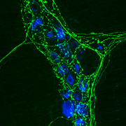
(see immunofluorescence
Immunofluorescence
Immunofluorescence is a technique used for light microscopy with a fluorescence microscope and is used primarily on biological samples. This technique uses the specificity of antibodies to their antigen to target fluorescent dyes to specific biomolecule targets within a cell, and therefore allows...
).
Sample preparation
While using the right antibodies to target the correct antigens and amplify the signal is important for visualization, complete preparation of the sample is critical to maintain cell morphology, tissue architecture and the antigenicity of target epitopes. This requires proper tissue collection, fixationFixation (histology)
In the fields of histology, pathology, and cell biology, fixation is a chemical process by which biological tissues are preserved from decay, thereby preventing autolysis or putrefaction...
and sectioning. Paraformaldehyde is usually used with fixation. Depending on the purpose and the thickness of the experimental sample, either thin (about 4-40 μm) sections are sliced from the tissue of interest, or if the tissue is not very thick and is penetrable it is used whole. The slicing is usually accomplished through the use of a microtome
Microtome
A microtome is a sectioning instrument that allows for the cutting of extremely thin slices of material, known as sections. Microtomes are an important device in microscopy preparation, allowing for the preparation of samples for observation under transmitted light or electron radiation...
, and slices are mounted on slides. "Free-floating IHC" uses slices that are not mounted, these slices are normally produced using a vibrating microtome
Microtome
A microtome is a sectioning instrument that allows for the cutting of extremely thin slices of material, known as sections. Microtomes are an important device in microscopy preparation, allowing for the preparation of samples for observation under transmitted light or electron radiation...
.
Because of the method of fixation and tissue preservation, the sample may require additional steps to make the epitopes available for antibody binding, including deparaffinization and antigen retrieval (microwave method, enzyme method, hot incubation method); these steps often makes the difference between staining and no staining. Additionally, depending on the tissue type and the method of antigen detection, endogenous biotin or enzymes may need to be blocked or quenched, respectively, prior to antibody staining.
Unlike immunocytochemistry
Immunocytochemistry
Immunocytochemistry is a common laboratory technique that uses antibodies that target specific peptides or protein antigens in the cell via specific epitopes. These bound antibodies can then be detected using several different methods. ICC allows researchers to evaluate whether or not cells in a...
, the tissue does not need to be permeabilized because this has already been accomplished by the microtome blade during sample preparation. Detergents like Triton X-100 are generally used in immunohistochemistry to reduce surface tension
Surface tension
Surface tension is a property of the surface of a liquid that allows it to resist an external force. It is revealed, for example, in floating of some objects on the surface of water, even though they are denser than water, and in the ability of some insects to run on the water surface...
, allowing less reagent to be used to achieve better and more even coverage of the sample.
Although antibodies show preferential avidity for specific epitopes, they may partially or weakly bind to sites on nonspecific proteins (also called reactive sites) that are similar to the cognate binding sites on the target antigen. In the context of antibody-mediated antigen detection, nonspecific binding causes high background staining that can mask the detection of the target antigen. To reduce background staining in IHC, ICC and any other immunostaining application, the samples are incubated with a buffer that blocks the reactive sites to which the primary or secondary antibodies may otherwise bind. Common blocking buffers include normal serum, non-fat dry milk, BSA or gelatin, and commercial blocking buffers with proprietary formulations are available for greater efficiency.
Antibody types
The antibodies used for specific detection can be polyclonal or monoclonalMonoclonal antibodies
Monoclonal antibodies are monospecific antibodies that are the same because they are made by identical immune cells that are all clones of a unique parent cell....
. Polyclonal antibodies are made by injecting animals with peptide Ag and, after a secondary immune response is stimulated, isolating antibodies from whole serum. Thus, polyclonal antibodies are a heterogeneous mix of antibodies that recognize several epitope
Epitope
An epitope, also known as antigenic determinant, is the part of an antigen that is recognized by the immune system, specifically by antibodies, B cells, or T cells. The part of an antibody that recognizes the epitope is called a paratope...
s. Monoclonal antibodies show specificity for a single epitope and are therefore considered more specific to the target antigen than polyclonal antibodies. For IHC detection strategies, antibodies are classified as primary or secondary reagents. Primary antibodies are raised against an antigen of interest and are typically unconjugated (unlabelled), while secondary antibodies are raised against immunoglobulins of the primary antibody species. The secondary antibody is usually conjugated to a linker molecule, such as biotin
Biotin
Biotin, also known as Vitamin H or Coenzyme R, is a water-soluble B-complex vitamin discovered by Bateman in 1916. It is composed of a ureido ring fused with a tetrahydrothiophene ring. A valeric acid substituent is attached to one of the carbon atoms of the tetrahydrothiophene ring...
, that then recruits reporter molecules, or the secondary antibody is directly bound to the reporter molecule itself.
IHC reporters
Reporter molecules vary based on the nature of the detection method, and the most popular methods of detection are with enzyme- and fluorophore-mediated chromogenic and fluorescenceFluorescence
Fluorescence is the emission of light by a substance that has absorbed light or other electromagnetic radiation of a different wavelength. It is a form of luminescence. In most cases, emitted light has a longer wavelength, and therefore lower energy, than the absorbed radiation...
detection, respectively. With chromogenic reporters, an enzyme label is reacted with a substrate to yield an intensely colored product that can be analyzed with an ordinary light microscope. While the list of enzyme substrates is extensive, Alkaline phosphatase
Alkaline phosphatase
Alkaline phosphatase is a hydrolase enzyme responsible for removing phosphate groups from many types of molecules, including nucleotides, proteins, and alkaloids. The process of removing the phosphate group is called dephosphorylation...
(AP) and horseradish peroxidase
Horseradish peroxidase
The enzyme horseradish peroxidase , found in horseradish, is used extensively in biochemistry applications primarily for its ability to amplify a weak signal and increase detectability of a target molecule.-Applications:...
(HRP) are the two enzymes used most extensively as labels for protein detection. An array of chromogenic, fluorogenic and chemiluminescent substrates is available for use with either enzyme, including DAB or BCIP/NBT
Nitro blue tetrazolium chloride
Nitro blue tetrazolium is a chemical compound composed of two tetrazole moieties. It is used in immunology for sensitive detection of alkaline phosphatase . NBT serves as the oxidant and BCIP is the AP-substrate ....
, which produce a brown or purple staining, respectively, wherever the enzymes are bound. Reaction with DAB can be enhanced using nickel
Nickel
Nickel is a chemical element with the chemical symbol Ni and atomic number 28. It is a silvery-white lustrous metal with a slight golden tinge. Nickel belongs to the transition metals and is hard and ductile...
, producing a deep purple/black staining. Fluorescent reporters are small, organic molecules used for IHC detection and traditionally include FITC
Fluorescein isothiocyanate
Fluorescein isothiocyanate is a derivative of fluorescein used in wide-ranging applications including flow cytometry. FITC is the original fluorescein molecule functionalized with an isothiocyanate reactive group , replacing a hydrogen atom on the bottom ring of the structure...
, TRITC
Rhodamine
Rhodamine is a family of related chemical compounds, fluorone dyes. Examples are Rhodamine 6G and Rhodamine B. They are used as a dye and as a dye laser gain medium. They are often used as a tracer dye within water to determine the rate and direction of flow and transport...
and AMCA, while commercial derivatives, including the Alexa Fluors and Dylight Fluors, show similar enhanced performance but vary in price. For chromogenic and fluorescent detection methods, densitometric analysis of the signal can provide semi- and fully quantitative data, respectively, to correlate the level of reporter signal to the level of protein expression or localization.
Target antigen detection methods
The direct method is a one-step staining method and involves a labeled antibodyAntibody
An antibody, also known as an immunoglobulin, is a large Y-shaped protein used by the immune system to identify and neutralize foreign objects such as bacteria and viruses. The antibody recognizes a unique part of the foreign target, termed an antigen...
(e.g. FITC
Fluorescein isothiocyanate
Fluorescein isothiocyanate is a derivative of fluorescein used in wide-ranging applications including flow cytometry. FITC is the original fluorescein molecule functionalized with an isothiocyanate reactive group , replacing a hydrogen atom on the bottom ring of the structure...
-conjugated antiserum
Antiserum
Antiserum is blood serum containing polyclonal antibodies. Antiserum is used to pass on passive immunity to many diseases. Passive antibody transfusion from a previous human survivor is the only known effective treatment for Ebola infection .The most common use of antiserum in humans is as...
) reacting directly with the antigen
Antigen
An antigen is a foreign molecule that, when introduced into the body, triggers the production of an antibody by the immune system. The immune system will then kill or neutralize the antigen that is recognized as a foreign and potentially harmful invader. These invaders can be molecules such as...
in tissue sections. While this technique utilizes only one antibody
Antibody
An antibody, also known as an immunoglobulin, is a large Y-shaped protein used by the immune system to identify and neutralize foreign objects such as bacteria and viruses. The antibody recognizes a unique part of the foreign target, termed an antigen...
and therefore is simple and rapid, the sensitivity is lower due to little signal amplification, such as with indirect methods, and is less commonly used than indirect methods.
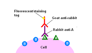
The indirect method involves an unlabeled primary antibody (first layer) that binds to the target antigen
Antigen
An antigen is a foreign molecule that, when introduced into the body, triggers the production of an antibody by the immune system. The immune system will then kill or neutralize the antigen that is recognized as a foreign and potentially harmful invader. These invaders can be molecules such as...
in the tissue and a labeled secondary antibody (second layer) that reacts with the primary antibody. As mentioned above, the secondary antibody must be raised against the IgG of the animal species in which the primary antibody has been raised. This method is more sensitive than direct detection strategies because of signal amplification due to the binding of several secondary antibodies to each primary antibody if the secondary antibody is conjugated to the fluorescent or enzyme
Enzyme
Enzymes are proteins that catalyze chemical reactions. In enzymatic reactions, the molecules at the beginning of the process, called substrates, are converted into different molecules, called products. Almost all chemical reactions in a biological cell need enzymes in order to occur at rates...
reporter. Further amplification can be achieved if the secondary antibody is conjugated to several biotin
Biotin
Biotin, also known as Vitamin H or Coenzyme R, is a water-soluble B-complex vitamin discovered by Bateman in 1916. It is composed of a ureido ring fused with a tetrahydrothiophene ring. A valeric acid substituent is attached to one of the carbon atoms of the tetrahydrothiophene ring...
molecules, which can recruit complexes of avidin
Avidin
Avidin is a tetrameric biotin-binding protein produced in the oviducts of birds, reptiles and amphibians deposited in the whites of their eggs. In chicken egg white, avidin makes up approximately 0.05% of total protein...
-, streptavidin
Streptavidin
Streptavidin is a 60000 dalton protein purified from the bacterium Streptomyces avidinii. Streptavidin homo-tetramers have an extraordinarily high affinity for biotin . With a dissociation constant on the order of ≈10-14 mol/L, the binding of biotin to streptavidin is one of the strongest...
or NeutrAvidin protein
NeutrAvidin
NeutrAvidin protein is a deglycosylated version of avidin, with a mass of approximately 60,000 daltons. As a result of carbohydrate removal, lectin binding is reduced to undetectable levels, yet biotin binding affinity is retained because the carbohydrate is not necessary for this activity...
bound-enzyme. The difference between these three biotin-binding proteins is their individual binding affinity to endogenous tissue targets leading to nonspecific binding and high background; the ranking of these proteins based on their nonspecific binding affinities, from highest to lowest, is: 1) avidin, 2) streptavidin and 3) Neutravidin protein.
The indirect method, aside from its greater sensitivity, also has the advantage that only a relatively small number of standard conjugated (labeled) secondary antibodies needs to be generated. For example, a labeled secondary antibody raised against rabbit IgG, which can be purchased "off the shelf," is useful with any primary antibody raised in rabbit. With the direct method, it would be necessary to label each primary antibody for every antigen of interest.
Counterstains
After immunohistochemical staining of the target antigen, a second stain is often applied to provide contrast that helps the primary stain stand out. Many of these stains show specificity for discrete cellular compartments or antigens, while others will stain the whole cell. Both chromogenic and fluorescent dyes are available for IHC to provide a vast array of reagents to fit every experimental design, and include: hematoxylin, Hoechst stainHoechst stain
Hoechst stains are part of a family of blue fluorescent dyes used to stain DNA. These Bis-benzimides were originally developed by the Hoechst AG, which numbered all their compounds so that the dye Hoechst 33342 is the 33342nd compound made by the company. There are three related Hoechst stains:...
and DAPI
DAPI
DAPI or 4',6-diamidino-2-phenylindole is a fluorescent stain that binds strongly to A-T rich regions in DNA. It is used extensively in fluorescence microscopy...
are commonly used.
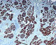
IHC Troubleshooting
In immunohistochemical techniques, there are several steps prior to the final staining of the tissue antigen, and many potential problems affect the outcome of the procedure. The major problem areas in IHC staining include strong background staining, weak target antigen staining and autofluorescence. Endogenous biotin or reporter enzymes or primary/secondary antibody cross-reactivity are common causes of strong background staining, while weak staining may be caused by poor enzyme activity or primary antibody potency. Furthermore, autofluorescence may be due to the nature of the tissue or the fixation method. These aspects of IHC tissue prep and antibody staining must be systematically addressed to identify and overcome staining issues.Diagnostic IHC markers
Western blot
The western blot is a widely used analytical technique used to detect specific proteins in the given sample of tissue homogenate or extract. It uses gel electrophoresis to separate native proteins by 3-D structure or denatured proteins by the length of the polypeptide...
or similar procedure. The technique is even more widely used in diagnostic surgical pathology
Surgical pathology
Surgical pathology is the most significant and time-consuming area of practice for most anatomical pathologists. Surgical pathology involves the gross and microscopic examination of surgical specimens, as well as biopsies submitted by non-surgeons such as general internists, medical subspecialists,...
for typing tumors (e.g. immunostaining for e-cadherin to differentiate between DCIS (ductal carcinoma in situ: stains positive) and LCIS (lobular carcinoma in situ: does not stain positive)).
- Carcinoembryonic antigenCarcinoembryonic antigenCarcinoembryonic antigen is a glycoprotein involved in cell adhesion. It is normally produced during fetal development, but the production of CEA stops before birth. Therefore, it is not usually present in the blood of healthy adults, although levels are raised in heavy smokers...
(CEA): used for identification of adenocarcinomaAdenocarcinomaAdenocarcinoma is a cancer of an epithelium that originates in glandular tissue. Epithelial tissue includes, but is not limited to, the surface layer of skin, glands and a variety of other tissue that lines the cavities and organs of the body. Epithelium can be derived embryologically from...
s. Not specific for site. - CytokeratinCytokeratinCytokeratins are proteins of keratin-containing intermediate filaments found in the intracytoplasmic cytoskeleton of epithelial tissue. The term "cytokeratin" began to be used in the late 1970s when the protein subunits of keratin intermediate filaments inside cells were first being identified and...
s: used for identification of carcinomas but may also be expressed in some sarcomas. - CD15Cluster of differentiationThe cluster of differentiation is a protocol used for the identification and investigation of cell surface molecules present on white blood cells, providing targets for immunophenotyping of cells...
and CD30 : used for Hodgkin's disease - Alpha fetoprotein: for yolk sac tumors and hepatocellular carcinomaHepatocellular carcinomaHepatocellular carcinoma is the most common type of liver cancer. Most cases of HCC are secondary to either a viral hepatitide infection or cirrhosis .Compared to other cancers, HCC is quite a rare tumor in the United States...
- CD117CD117Mast/stem cell growth factor receptor also known as proto-oncogene c-Kit or tyrosine-protein kinase Kit or CD117 is a protein that in humans is encoded by the KIT gene...
(KIT): for gastrointestinal stromal tumorGastrointestinal stromal tumorA gastrointestinal stromal tumor is one of the most common mesenchymal tumors of the gastrointestinal tract...
s (GIST) - CD10 (CALLA): for renal cell carcinomaRenal cell carcinomaRenal cell carcinoma is a kidney cancer that originates in the lining of the proximal convoluted tubule, the very small tubes in the kidney that filter the blood and remove waste products. RCC is the most common type of kidney cancer in adults, responsible for approximately 80% of cases...
and acute lymphoblastic leukemiaAcute lymphoblastic leukemiaAcute lymphoblastic leukemia is a form of leukemia, or cancer of the white blood cells characterized by excess lymphoblasts.Malignant, immature white blood cells continuously multiply and are overproduced in the bone marrow. ALL causes damage and death by crowding out normal cells in the bone... - Prostate specific antigenProstate specific antigenProstate-specific antigen also known as gamma-seminoprotein or kallikrein-3 is a glycoprotein that in humans is encoded by the KLK3 gene. KLK3 is a member of the kallikrein-related peptidase family that are secreted by the epithelial cells of the prostate gland...
(PSA): for prostate cancerProstate cancerProstate cancer is a form of cancer that develops in the prostate, a gland in the male reproductive system. Most prostate cancers are slow growing; however, there are cases of aggressive prostate cancers. The cancer cells may metastasize from the prostate to other parts of the body, particularly... - estrogens and progesteroneProgesteroneProgesterone also known as P4 is a C-21 steroid hormone involved in the female menstrual cycle, pregnancy and embryogenesis of humans and other species...
staining for tumour identification - Identification of B-cell lymphomas using CD20CD20B-lymphocyte antigen CD20 or CD20 is an activated-glycosylated phosphoprotein expressed on the surface of all B-cells beginning at the pro-B phase and progressively increasing in concentration until maturity....
- Identification of T-cell lymphomas using CD3CD3CD3 or CD-3 may be:* CD3 , an antigen, cluster of differentiation protein , part of the T cell receptor complex on a mature T lymphocyte* Ford CD3 platform* MediaMax CD-3, copy protection scheme* MiniCD, a 3-inch CD...
Directing therapy
A variety of molecular pathways are altered in cancer and some of the alterations can be targeted in cancer therapy. Immunohistochemistry can be used to assess which tumors are likely to respond to therapy, by detecting the presence or elevated levels of the molecular target.Chemical inhibitors
Tumor biology allows for a number of potential intracellular targets. Many tumors are hormone dependent. The presence of hormone receptors can be used to determine if a tumor is potentially responsive to antihormonal therapy. One of the first therapies was the antiestrogen, tamoxifenTamoxifen
Tamoxifen is an antagonist of the estrogen receptor in breast tissue via its active metabolite, hydroxytamoxifen. In other tissues such as the endometrium, it behaves as an agonist, hence tamoxifen may be characterized as a mixed agonist/antagonist...
, used to treat breast cancer. Such hormone receptors can be detected by immunohistochemistry.
Imatinib
Imatinib
Imatinib is a drug used to treat certain types of cancer. It is currently marketed by Novartis as Gleevec or Glivec as its mesylate salt, imatinib mesilate . It is used in treating chronic myelogenous leukemia , gastrointestinal stromal tumors and some other diseases...
, an intracellualar tyrosine kinase
Tyrosine kinase
A tyrosine kinase is an enzyme that can transfer a phosphate group from ATP to a protein in a cell. It functions as an "on" or "off" switch in many cellular functions....
inhibitor, was developed to treat chronic myelogenous leukemia
Chronic myelogenous leukemia
Chronic myelogenous leukemia , also known as chronic granulocytic leukemia , is a cancer of the white blood cells. It is a form of leukemia characterized by the increased and unregulated growth of predominantly myeloid cells in the bone marrow and the accumulation of these cells in the blood...
, a disease characterized by the formation of a specific abnormal tyrosine kinase. Imitanib has proven effective in tumors, that express other tyrosine kinases, most notably KIT. Most gastrointestinal stromal tumor
Gastrointestinal stromal tumor
A gastrointestinal stromal tumor is one of the most common mesenchymal tumors of the gastrointestinal tract...
s express KIT, which can be detected by immunohistochemistry.
Monoclonal antibodies
Many proteins shown to be highly upregulated in pathological states by immunohistochemistry are potential targets for therapies utilising monoclonal antibodies. Monoclonal antibodies, due to their size, are utilized against cell surface targets. Among the overexpressed targets, the members of the epidermal growth factor receptorEpidermal growth factor receptor
The epidermal growth factor receptor is the cell-surface receptor for members of the epidermal growth factor family of extracellular protein ligands...
(EGFR) family, transmembrane proteins with an extracellular receptor domain regulating an intracellular tyrosine kinase, Of these, HER2/neu
HER2/neu
HER-2 also known as proto-oncogene Neu, receptor tyrosine-protein kinase erbB-2, CD340 or p185 is an enzyme that in humans is encoded by the ERBB2 gene. Over expression of this gene is correlated with higher aggressiveness in breast cancers...
(also known as Erb-B2) was the first to be developed. The molecule is highly expressed in a variety of cancer cell types, most notably breast cancer. As such, antibodies against HER2/neu have been FDA approved for clinical treatment of cancer under the drug name Herceptin. There are commercially available immunohistochemical tests, Dako HercepTest and Ventana Pathway.
Similarly, EGFR (HER-1) is overexpressed in a variety of cancers including head and neck and colon. Immunohistochemistry is used to determine patients who may benefit from therapeutic antibodies such as Erbitux (cetuximab). Commercial systems to detect EGFR by immunohistochemistry include the Dako pharmDx.
External links
- Overview of Immunohistochemistry--describes all aspects of IHC including sample prep, staining and troubleshooting
- Yale Core Tissue Microarray Facility
- Histochemical Staining Methods - University of RochesterUniversity of RochesterThe University of Rochester is a private, nonsectarian, research university in Rochester, New York, United States. The university grants undergraduate and graduate degrees, including doctoral and professional degrees. The university has six schools and various interdisciplinary programs.The...
Department of Pathology - Immunohistochemistry Staining Protocol
- HistoWiki entry for Immunohistochemistry