L-isoaspartyl methyltransferase
Encyclopedia
Protein L-isoaspartyl methyltransferase (PIMT, PCMT), also called S-adenosyl-L-methionine:protein-L-isoaspartate O-methyltransferase, is an enzyme
which recognizes and catalyzes the repair of damaged L-isoaspartyl
and D-aspartatyl
groups in proteins. It is a highly conserved enzyme which is present in nearly all eukaryotes, archaebacteria, and Gram-negative
eubacteria.
side chain and adds it to the side chain carboxyl group of L-isoaspartate or D-aspartate to create a methyl ester. Subsequent nonenzymatic reactions result in a rapid transformation to L-succinimide, which is a precursor to aspartate and isoaspartate. The L-succinimide can then undergo nonenzymatic hydrolysis, which generates some repaired L-aspartyl residues as well as some L-isoaspartyl residues, which can then enter the cycle again for eventual conversion to the normal peptide linkage.
PIMT tends to act on proteins that have been non-enzymatically damaged due to age. By performing this repair mechanism, the enzyme helps to maintain overall protein integrity. This mechanism has been observed by several groups, and has been confirmed through experimental testing. In one report, PIMT was inhibited by adenosine dialdehyde. The results supported the proposed function of the enzyme, as the amount of abnormal L-aspartate residues increased when cells were treated with the indirect inhibitor, adenosine dialdehyde. Additionally, S-adenosylhomocysteine is known to be a competitive inhibitor of PIMT. When PIMT is not present in cells, the abnormal aspartyl residues accumulate, creating abnormal proteins that have been known to cause fatal progressive epilepsy
in mice. It has been suggested that calmodulin
may play a role in stimulating the function of PIMT, although the relationship between these two molecules has not been thoroughly explored. In addition to calmodulin, guanosine 5'-O-[gamma-thio]triphosphate (GTPgammaS
) has been found to stimulate PIMT activity.
in two forms due to alternative splicing
and differs among individuals in the population due to a single polymorphism
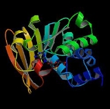
at protein 119, either valine or isoleucine. The enzyme structure is described as a “doubly wound alpha/beta/alpha sandwich structure” which is quite consistent in all species analyzed thus far. If there is any difference in the sequences between different organisms it occurs in the regions connecting the three motifs in the sandwich structure, but the sequence of the individual motifs tends to be highly conserved. Researchers have found the active site
to be in the loop between the beta structure and the second alpha helix and have determined it to be highly specific for isoaptartyl residues. For example, the residues found at the C-terminus of drosophila
PIMT (dPIMT) are rotated 90 degrees so as to allow more space for a substrate to interact with the enzyme. In fact, dPIMT appears to alternate between this unique open conformation and the less open conformation common of PIMT in other organisms. Although possibly unrelated to this, increased levels of dPIMT in drosophila have been correlated with increase life expectancy in these organisms due to their importance in protein repair.
Enzyme
Enzymes are proteins that catalyze chemical reactions. In enzymatic reactions, the molecules at the beginning of the process, called substrates, are converted into different molecules, called products. Almost all chemical reactions in a biological cell need enzymes in order to occur at rates...
which recognizes and catalyzes the repair of damaged L-isoaspartyl
Isoaspartate
The isoaspartate group is a functional group in biochemistry. Its formation is a chemical reaction in which the side chain of an asparagine or aspartic acid residue attacks the following peptide group , forming a symmetric succinimide intermediate...
and D-aspartatyl
Aspartic acid
Aspartic acid is an α-amino acid with the chemical formula HOOCCHCH2COOH. The carboxylate anion, salt, or ester of aspartic acid is known as aspartate. The L-isomer of aspartate is one of the 20 proteinogenic amino acids, i.e., the building blocks of proteins...
groups in proteins. It is a highly conserved enzyme which is present in nearly all eukaryotes, archaebacteria, and Gram-negative
Gram-negative
Gram-negative bacteria are bacteria that do not retain crystal violet dye in the Gram staining protocol. In a Gram stain test, a counterstain is added after the crystal violet, coloring all Gram-negative bacteria with a red or pink color...
eubacteria.
Function
PIMT acts to transfer methyl groups from S-adenosyl-L-methionine to the alpha side chain carboxyl groups of damaged L-isoaspartyl and D-aspartatyl amino acids. The enzyme takes the end methyl residue from the methionineMethionine
Methionine is an α-amino acid with the chemical formula HO2CCHCH2CH2SCH3. This essential amino acid is classified as nonpolar. This amino-acid is coded by the codon AUG, also known as the initiation codon, since it indicates mRNA's coding region where translation into protein...
side chain and adds it to the side chain carboxyl group of L-isoaspartate or D-aspartate to create a methyl ester. Subsequent nonenzymatic reactions result in a rapid transformation to L-succinimide, which is a precursor to aspartate and isoaspartate. The L-succinimide can then undergo nonenzymatic hydrolysis, which generates some repaired L-aspartyl residues as well as some L-isoaspartyl residues, which can then enter the cycle again for eventual conversion to the normal peptide linkage.
PIMT tends to act on proteins that have been non-enzymatically damaged due to age. By performing this repair mechanism, the enzyme helps to maintain overall protein integrity. This mechanism has been observed by several groups, and has been confirmed through experimental testing. In one report, PIMT was inhibited by adenosine dialdehyde. The results supported the proposed function of the enzyme, as the amount of abnormal L-aspartate residues increased when cells were treated with the indirect inhibitor, adenosine dialdehyde. Additionally, S-adenosylhomocysteine is known to be a competitive inhibitor of PIMT. When PIMT is not present in cells, the abnormal aspartyl residues accumulate, creating abnormal proteins that have been known to cause fatal progressive epilepsy
Epilepsy
Epilepsy is a common chronic neurological disorder characterized by seizures. These seizures are transient signs and/or symptoms of abnormal, excessive or hypersynchronous neuronal activity in the brain.About 50 million people worldwide have epilepsy, and nearly two out of every three new cases...
in mice. It has been suggested that calmodulin
Calmodulin
Calmodulin is a calcium-binding protein expressed in all eukaryotic cells...
may play a role in stimulating the function of PIMT, although the relationship between these two molecules has not been thoroughly explored. In addition to calmodulin, guanosine 5'-O-[gamma-thio]triphosphate (GTPgammaS
GTPgammaS
GTPgammaS is nonhydrolyzable G-protein-activating analog of guanosine triphosphate...
) has been found to stimulate PIMT activity.
Structure
The enzyme is present in human cytosolCytosol
The cytosol or intracellular fluid is the liquid found inside cells, that is separated into compartments by membranes. For example, the mitochondrial matrix separates the mitochondrion into compartments....
in two forms due to alternative splicing
Alternative splicing
Alternative splicing is a process by which the exons of the RNA produced by transcription of a gene are reconnected in multiple ways during RNA splicing...
and differs among individuals in the population due to a single polymorphism
Polymorphism (biology)
Polymorphism in biology occurs when two or more clearly different phenotypes exist in the same population of a species — in other words, the occurrence of more than one form or morph...
at protein 119, either valine or isoleucine. The enzyme structure is described as a “doubly wound alpha/beta/alpha sandwich structure” which is quite consistent in all species analyzed thus far. If there is any difference in the sequences between different organisms it occurs in the regions connecting the three motifs in the sandwich structure, but the sequence of the individual motifs tends to be highly conserved. Researchers have found the active site
Active site
In biology the active site is part of an enzyme where substrates bind and undergo a chemical reaction. The majority of enzymes are proteins but RNA enzymes called ribozymes also exist. The active site of an enzyme is usually found in a cleft or pocket that is lined by amino acid residues that...
to be in the loop between the beta structure and the second alpha helix and have determined it to be highly specific for isoaptartyl residues. For example, the residues found at the C-terminus of drosophila
Drosophila
Drosophila is a genus of small flies, belonging to the family Drosophilidae, whose members are often called "fruit flies" or more appropriately pomace flies, vinegar flies, or wine flies, a reference to the characteristic of many species to linger around overripe or rotting fruit...
PIMT (dPIMT) are rotated 90 degrees so as to allow more space for a substrate to interact with the enzyme. In fact, dPIMT appears to alternate between this unique open conformation and the less open conformation common of PIMT in other organisms. Although possibly unrelated to this, increased levels of dPIMT in drosophila have been correlated with increase life expectancy in these organisms due to their importance in protein repair.

