Lateral geniculate nucleus
Encyclopedia
The lateral geniculate nucleus (LGN) is the primary relay center for visual
information received from the retina
of the eye
. The LGN is found inside the thalamus
of the brain
.
The LGN receives information directly from the ascending retinal ganglion cells via the optic tract
and from the reticular activating system
. Neurons of the LGN send their axons through the optic radiation
, a pathway directly to the primary visual cortex. In addition, the LGN receives many strong feedback connections from the primary visual cortex. In mammals and humans the two strongest pathways linking the eye to the brain are those projecting to the LGNd (dorsal part of the LGN in the thalamus), and to the Superior Colliculus (SC)
of the brain have a lateral geniculate nucleus, named so for its resemblance to a bent knee (genu is Latin for "knee"). In many primate
s, including humans and macaque
s, it has layers of cell bodies with layers of neuropil
in between, in an arrangement something like a club sandwich
or layer cake
, with cell bodies of LGN neuron
s as the "cake" and neuropil
as the "icing
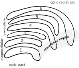
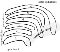
". In humans and macaques the LGN is normally described as having six distinctive layers. The inner two layers, 1 and 2, are called the magnocellular layers, while the outer four layers, 3, 4, 5, and 6, are called parvocellular layers. An additional set of neurons, known as the koniocellular sublayers, are found ventral to each of the magnocellular and parvocellular layers. It must be noted, this layering is variable between primate species, and extra leafleting is variable within species.

The magnocellular, parvocellular, and koniocellular layers of the LGN correspond with the similarly named types of ganglion cell
s.
Koniocellular cells are functionally and neurochemically distinct from M and P cells and provide a third channel to the visual cortex. They project their axons between the layers of the lateral geniculate nucleus where M and P cells project. Their role in visual perception is presently unclear; however, the koniocellular system has been linked with the integration of somatosensory system-proprioceptive information with visual perception, and it may also be involved in color perception.
The parvo- and magnocellular fibers were previously thought to dominate the Ungerleider-Mishkin ventral stream and dorsal stream, respectively. However, new evidence has accumulated showing that the two streams appear to feed on a more even mixture of different types of nerve fibers.
The other major retino-cortical visual pathway is the retinotectal pathway, routing primarily through the superior colliculus
and thalamic pulvinar
nucleus onto posterior parietal cortex and visual area MT.
Within one LGN, the visual information is divided among the various layers as follows:
A simple mnemonic
for remembering this is "See I? I see, I see," with "see" representing the C in "contralateral," and "I" representing the I in "ipsilateral."
Another way of remembering this is 2+3=5, which is correct, so ipsilateral side, and 1+4 doesn't equal 6, so contralateral.
This description applies to the LGN of many primates, but not all. The sequence of layers receiving information from the ipsilateral and contralateral (opposite side of the head) eyes is different in the tarsier
. Some neuroscientists suggested that "this apparent difference distinguishes tarsiers from all other primates, reinforcing the view that they arose in an early, independent line of primate evolution".
In visual perception
, the right eye gets information from the right side of the world (the right visual field
), as well as the left side of the world (the left visual field
). You can confirm this by covering your left eye: the right eye still sees to your left and right, although on the left side your field of view is partially blocked by your nose.
In the LGN, the corresponding information from the right and left eyes is "stacked" so that a toothpick
driven through the club sandwich
of layers 1 through 6 would hit the same point in visual space six different times.
At least in some species, the LGN also receives some inputs from the optic tectum (also known as the superior colliculus
).
s, which form part of the retrolenticular limb of the internal capsule
.
The axon
s that leave the LGN go to V1 visual cortex
. Both the magnocellular layers 1-2 and the parvocellular layers 3-6 send their axons to layer 4 in V1. Within layer 4 of V1, layer 4cβ receives parvocellular input, and layer 4cα receives magnocellular input. However, the koniocellular layers (in between layers 1-6) send their axons to layers 4a in V1.
Axon
s from layer 6 of visual cortex
send information back to the LGN.
Studies involving blindsight
have suggested that projections from the LGN not only travel to the primary visual cortex but also to higher cortical areas V2 and V3. Patients with blindsight are phenomenally blind in certain areas of the visual field corresponding to a contralateral lesion in primary visual cortex; however, these patients are able to perform certain motor tasks accurately in their blind field, such as grasping. This suggests that neurons travel from the LGN to both the visual cortex and higher cortex regions.
through center surround inhibition, the LGN accomplishes temporal decorrelation. This spatial-temporal decorrelation makes for much more efficient coding. However, there is almost certainly much more going on.
Like other areas of the thalamus
, particularly other relay nuclei, the LGN likely helps the visual system
focus its attention on the most important information. That is, if you hear a sound slightly to your left, the auditory system
likely "tells" the visual system
, through the LGN, to direct visual attention to that part of space.
The LGN is also a station that refines certain receptive fields.
Recent experiments using fMRI in humans have found that both spatial attention and saccadic eye movement
s can modulate activity in the LGN.
Visual perception
Visual perception is the ability to interpret information and surroundings from the effects of visible light reaching the eye. The resulting perception is also known as eyesight, sight, or vision...
information received from the retina
Retina
The vertebrate retina is a light-sensitive tissue lining the inner surface of the eye. The optics of the eye create an image of the visual world on the retina, which serves much the same function as the film in a camera. Light striking the retina initiates a cascade of chemical and electrical...
of the eye
Human eye
The human eye is an organ which reacts to light for several purposes. As a conscious sense organ, the eye allows vision. Rod and cone cells in the retina allow conscious light perception and vision including color differentiation and the perception of depth...
. The LGN is found inside the thalamus
Thalamus
The thalamus is a midline paired symmetrical structure within the brains of vertebrates, including humans. It is situated between the cerebral cortex and midbrain, both in terms of location and neurological connections...
of the brain
Human brain
The human brain has the same general structure as the brains of other mammals, but is over three times larger than the brain of a typical mammal with an equivalent body size. Estimates for the number of neurons in the human brain range from 80 to 120 billion...
.
The LGN receives information directly from the ascending retinal ganglion cells via the optic tract
Optic tract
The optic tract is a part of the visual system in the brain.It is a continuation of the optic nerve and runs from the optic chiasm to the lateral geniculate nucleus....
and from the reticular activating system
Reticular activating system
The reticular activating system is an area of the brain responsible for regulating arousal and sleep-wake transitions.- History and Etymology :...
. Neurons of the LGN send their axons through the optic radiation
Optic radiation
The optic radiation is a collection of axons from relay neurons in the lateral geniculate nucleus of the thalamus carrying visual information to the visual cortex along the calcarine fissure.There is one such tract on each side of the brain.-Parts:A distinctive...
, a pathway directly to the primary visual cortex. In addition, the LGN receives many strong feedback connections from the primary visual cortex. In mammals and humans the two strongest pathways linking the eye to the brain are those projecting to the LGNd (dorsal part of the LGN in the thalamus), and to the Superior Colliculus (SC)
Structure
Both the left and right hemisphereCerebral hemisphere
A cerebral hemisphere is one of the two regions of the eutherian brain that are delineated by the median plane, . The brain can thus be described as being divided into left and right cerebral hemispheres. Each of these hemispheres has an outer layer of grey matter called the cerebral cortex that is...
of the brain have a lateral geniculate nucleus, named so for its resemblance to a bent knee (genu is Latin for "knee"). In many primate
Primate
A primate is a mammal of the order Primates , which contains prosimians and simians. Primates arose from ancestors that lived in the trees of tropical forests; many primate characteristics represent adaptations to life in this challenging three-dimensional environment...
s, including humans and macaque
Macaque
The macaques constitute a genus of Old World monkeys of the subfamily Cercopithecinae. - Description :Aside from humans , the macaques are the most widespread primate genus, ranging from Japan to Afghanistan and, in the case of the barbary macaque, to North Africa...
s, it has layers of cell bodies with layers of neuropil
Neuropil
In neuroanatomy, a neuropil, which is sometimes referred to as a neuropile, is a region between neuronal cell bodies in the gray matter of the brain and blood-brain barrier . It consists of a dense tangle of axon terminals, dendrites and glial cell processes...
in between, in an arrangement something like a club sandwich
Club sandwich
A club sandwich, also called a clubhouse sandwich or double-decker, is a sandwich with two layers of fillings between 3 slices of toasted bread. It is often cut into quarters and held together by hors d'œuvre sticks....
or layer cake
Cake
Cake is a form of bread or bread-like food. In its modern forms, it is typically a sweet and enriched baked dessert. In its oldest forms, cakes were normally fried breads or cheesecakes, and normally had a disk shape...
, with cell bodies of LGN neuron
Neuron
A neuron is an electrically excitable cell that processes and transmits information by electrical and chemical signaling. Chemical signaling occurs via synapses, specialized connections with other cells. Neurons connect to each other to form networks. Neurons are the core components of the nervous...
s as the "cake" and neuropil
Neuropil
In neuroanatomy, a neuropil, which is sometimes referred to as a neuropile, is a region between neuronal cell bodies in the gray matter of the brain and blood-brain barrier . It consists of a dense tangle of axon terminals, dendrites and glial cell processes...
as the "icing
Icing (food)
Icing, also called frosting in the United States, is a sweet often creamy glaze made of sugar with a liquid such as water or milk, that is often enriched with ingredients such as butter, egg whites, cream cheese, or flavorings and is used to cover or decorate baked goods, such as cakes or cookies...
". In humans and macaques the LGN is normally described as having six distinctive layers. The inner two layers, 1 and 2, are called the magnocellular layers, while the outer four layers, 3, 4, 5, and 6, are called parvocellular layers. An additional set of neurons, known as the koniocellular sublayers, are found ventral to each of the magnocellular and parvocellular layers. It must be noted, this layering is variable between primate species, and extra leafleting is variable within species.
M, P, K cells
| Type | Size* | Source / Type of Information | Location | Response | Number >- | M: Magnocellular cells |
Large | Rods; necessary for the perception of movement, depth, and small differences in brightness | Layers 1 and 2 | rapid and transient | >- | Small | Cones; long- and medium-wavelength ("red" and "green" cones); necessary for the perception of color and form (fine details). | Layers 3, 4, 5 and 6 | slow and sustained | >- | Very small cell bodies | Short-wavelength "blue" cones. | Between each of the M and P layers | ? |

- Size relates to cell body, dendritic tree and receptive field
The magnocellular, parvocellular, and koniocellular layers of the LGN correspond with the similarly named types of ganglion cell
Ganglion cell
A retinal ganglion cell is a type of neuron located near the inner surface of the retina of the eye. It receives visual information from photoreceptors via two intermediate neuron types: bipolar cells and amacrine cells...
s.
Koniocellular cells are functionally and neurochemically distinct from M and P cells and provide a third channel to the visual cortex. They project their axons between the layers of the lateral geniculate nucleus where M and P cells project. Their role in visual perception is presently unclear; however, the koniocellular system has been linked with the integration of somatosensory system-proprioceptive information with visual perception, and it may also be involved in color perception.
The parvo- and magnocellular fibers were previously thought to dominate the Ungerleider-Mishkin ventral stream and dorsal stream, respectively. However, new evidence has accumulated showing that the two streams appear to feed on a more even mixture of different types of nerve fibers.
The other major retino-cortical visual pathway is the retinotectal pathway, routing primarily through the superior colliculus
Superior colliculus
The optic tectum or simply tectum is a paired structure that forms a major component of the vertebrate midbrain. In mammals this structure is more commonly called the superior colliculus , but, even in mammals, the adjective tectal is commonly used. The tectum is a layered structure, with a...
and thalamic pulvinar
Pulvinar
The pulvinar nuclei are a collection of nuclei located in the pulvinar thalamus. The pulvinar part is the most posterior region of the thalamus....
nucleus onto posterior parietal cortex and visual area MT.
Ipsilateral and contralateral layers
Both the LGN in the right hemisphere and the LGN in the left hemisphere receive input from each eye. However, each LGN only receives information from one half of the visual field. This occurs due to axons of the ganglion cells from the inner halves of the retina (the nasal sides) decussating (crossing to the other side of the brain) through the optic chiasm (khiasma means "cross"). The axons of the ganglion cells from the outer half of the retina (the temporal sides) remain on the same side of the brain. Therefore, the right hemisphere receives visual information from the left visual field, and the left hemisphere receives visual information from the right visual field.Within one LGN, the visual information is divided among the various layers as follows:
- the eye on the same side (the ipsilateral eye) sends information to layers 2, 3 and 5
- the eye on the opposite side (the contralateral eye) sends information to layers 1, 4 and 6.
A simple mnemonic
Mnemonic
A mnemonic , or mnemonic device, is any learning technique that aids memory. To improve long term memory, mnemonic systems are used to make memorization easier. Commonly encountered mnemonics are often verbal, such as a very short poem or a special word used to help a person remember something,...
for remembering this is "See I? I see, I see," with "see" representing the C in "contralateral," and "I" representing the I in "ipsilateral."
Another way of remembering this is 2+3=5, which is correct, so ipsilateral side, and 1+4 doesn't equal 6, so contralateral.
This description applies to the LGN of many primates, but not all. The sequence of layers receiving information from the ipsilateral and contralateral (opposite side of the head) eyes is different in the tarsier
Tarsier
Tarsiers are haplorrhine primates of the genus Tarsius, a genus in the family Tarsiidae, which is itself the lone extant family within the infraorder Tarsiiformes...
. Some neuroscientists suggested that "this apparent difference distinguishes tarsiers from all other primates, reinforcing the view that they arose in an early, independent line of primate evolution".
In visual perception
Visual perception
Visual perception is the ability to interpret information and surroundings from the effects of visible light reaching the eye. The resulting perception is also known as eyesight, sight, or vision...
, the right eye gets information from the right side of the world (the right visual field
Visual field
The term visual field is sometimes used as a synonym to field of view, though they do not designate the same thing. The visual field is the "spatial array of visual sensations available to observation in introspectionist psychological experiments", while 'field of view' "refers to the physical...
), as well as the left side of the world (the left visual field
Visual field
The term visual field is sometimes used as a synonym to field of view, though they do not designate the same thing. The visual field is the "spatial array of visual sensations available to observation in introspectionist psychological experiments", while 'field of view' "refers to the physical...
). You can confirm this by covering your left eye: the right eye still sees to your left and right, although on the left side your field of view is partially blocked by your nose.
In the LGN, the corresponding information from the right and left eyes is "stacked" so that a toothpick
Toothpick
A toothpick is a small stick of wood, plastic, bamboo, metal, bone or other substance used to remove detritus from the teeth, usually after a meal. A toothpick usually has one or two sharp ends to insert between teeth. They can also be used for picking up small appetizers or as a cocktail...
driven through the club sandwich
Club sandwich
A club sandwich, also called a clubhouse sandwich or double-decker, is a sandwich with two layers of fillings between 3 slices of toasted bread. It is often cut into quarters and held together by hors d'œuvre sticks....
of layers 1 through 6 would hit the same point in visual space six different times.
LGN inputs
The LGN receives input from many sources, including the cortex and then sends its output to the cortex.At least in some species, the LGN also receives some inputs from the optic tectum (also known as the superior colliculus
Superior colliculus
The optic tectum or simply tectum is a paired structure that forms a major component of the vertebrate midbrain. In mammals this structure is more commonly called the superior colliculus , but, even in mammals, the adjective tectal is commonly used. The tectum is a layered structure, with a...
).
LGN output
Information leaving the LGN travels out on the optic radiationOptic radiation
The optic radiation is a collection of axons from relay neurons in the lateral geniculate nucleus of the thalamus carrying visual information to the visual cortex along the calcarine fissure.There is one such tract on each side of the brain.-Parts:A distinctive...
s, which form part of the retrolenticular limb of the internal capsule
Internal capsule
The internal capsule is an area of white matter in the brain that separates the caudate nucleus and the thalamus from the lenticular nucleus. The internal capsule contains both ascending and descending axons....
.
The axon
Axon
An axon is a long, slender projection of a nerve cell, or neuron, that conducts electrical impulses away from the neuron's cell body or soma....
s that leave the LGN go to V1 visual cortex
Visual cortex
The visual cortex of the brain is the part of the cerebral cortex responsible for processing visual information. It is located in the occipital lobe, in the back of the brain....
. Both the magnocellular layers 1-2 and the parvocellular layers 3-6 send their axons to layer 4 in V1. Within layer 4 of V1, layer 4cβ receives parvocellular input, and layer 4cα receives magnocellular input. However, the koniocellular layers (in between layers 1-6) send their axons to layers 4a in V1.
Axon
Axon
An axon is a long, slender projection of a nerve cell, or neuron, that conducts electrical impulses away from the neuron's cell body or soma....
s from layer 6 of visual cortex
Visual cortex
The visual cortex of the brain is the part of the cerebral cortex responsible for processing visual information. It is located in the occipital lobe, in the back of the brain....
send information back to the LGN.
Studies involving blindsight
Blindsight
Blindsight is a phenomenon in which people who are perceptually blind in a certain area of their visual field demonstrate some response to visual stimuli...
have suggested that projections from the LGN not only travel to the primary visual cortex but also to higher cortical areas V2 and V3. Patients with blindsight are phenomenally blind in certain areas of the visual field corresponding to a contralateral lesion in primary visual cortex; however, these patients are able to perform certain motor tasks accurately in their blind field, such as grasping. This suggests that neurons travel from the LGN to both the visual cortex and higher cortex regions.
Function in visual perception
The function of the LGN is unknown. It has been shown that while the retina accomplishes spatial decorrelationDecorrelation
Decorrelation is a general term for any process that is used to reduce autocorrelation within a signal, or cross-correlation within a set of signals, while preserving other aspects of the signal. A frequently used method of decorrelation is the use of a matched linear filter to reduce the...
through center surround inhibition, the LGN accomplishes temporal decorrelation. This spatial-temporal decorrelation makes for much more efficient coding. However, there is almost certainly much more going on.
Like other areas of the thalamus
Thalamus
The thalamus is a midline paired symmetrical structure within the brains of vertebrates, including humans. It is situated between the cerebral cortex and midbrain, both in terms of location and neurological connections...
, particularly other relay nuclei, the LGN likely helps the visual system
Visual system
The visual system is the part of the central nervous system which enables organisms to process visual detail, as well as enabling several non-image forming photoresponse functions. It interprets information from visible light to build a representation of the surrounding world...
focus its attention on the most important information. That is, if you hear a sound slightly to your left, the auditory system
Auditory system
The auditory system is the sensory system for the sense of hearing.- Outer ear :The folds of cartilage surrounding the ear canal are called the pinna...
likely "tells" the visual system
Visual system
The visual system is the part of the central nervous system which enables organisms to process visual detail, as well as enabling several non-image forming photoresponse functions. It interprets information from visible light to build a representation of the surrounding world...
, through the LGN, to direct visual attention to that part of space.
The LGN is also a station that refines certain receptive fields.
Recent experiments using fMRI in humans have found that both spatial attention and saccadic eye movement
Saccade
A saccade is a fast movement of an eye, head or other part of an animal's body or device. It can also be a fast shift in frequency of an emitted signal or other quick change. Saccades are quick, simultaneous movements of both eyes in the same direction...
s can modulate activity in the LGN.
External links
- Malpeli J. Malpeli Lab Home Page. Retrieved September 1, 2004. - "The Visual Pathway from Below"

