
Visual system
Encyclopedia
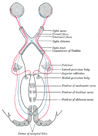
Central nervous system
The central nervous system is the part of the nervous system that integrates the information that it receives from, and coordinates the activity of, all parts of the bodies of bilaterian animals—that is, all multicellular animals except sponges and radially symmetric animals such as jellyfish...
which enables organisms to process visual detail
Visual perception
Visual perception is the ability to interpret information and surroundings from the effects of visible light reaching the eye. The resulting perception is also known as eyesight, sight, or vision...
, as well as enabling several non-image forming photoresponse functions. It interprets information from visible light to build a representation of the surrounding world. The visual system accomplishes a number of complex tasks, including the reception of light and the formation of monocular representations; the construction of a binocular perception from a pair of two dimensional projections; the identification and categorization of visual objects; assessing distances to and between objects; and guiding body movements in relation to visual objects. The psychological manifestation of visual information is known as visual perception
Visual perception
Visual perception is the ability to interpret information and surroundings from the effects of visible light reaching the eye. The resulting perception is also known as eyesight, sight, or vision...
, a lack of which is called blindness
Blindness
Blindness is the condition of lacking visual perception due to physiological or neurological factors.Various scales have been developed to describe the extent of vision loss and define blindness...
. Non-image forming visual functions, independent of visual perception, include the pupillary light reflex (PLR) and circadian photoentrainment
Entrainment (chronobiology)
Entrainment, within the study of chronobiology, occurs when rhythmic physiological or behavioral events match their period and phase to that of an environmental oscillation. A common example is the entrainment of circadian rhythms to the daily light–dark cycle, which ultimately is determined by...
.
Introduction
Mammal
Mammals are members of a class of air-breathing vertebrate animals characterised by the possession of endothermy, hair, three middle ear bones, and mammary glands functional in mothers with young...
s, although other "higher" animals have similar visual systems. In this case, the visual system consists of:
- The eye, especially the retina
- The optic nerve
- The optic chiasma
- The optic tract
- The lateral geniculate body
- The optic radiation
- The visual cortex
- The visual association cortex.
Different species
Species
In biology, a species is one of the basic units of biological classification and a taxonomic rank. A species is often defined as a group of organisms capable of interbreeding and producing fertile offspring. While in many cases this definition is adequate, more precise or differing measures are...
are able to see different parts of the light spectrum; for example, bee
Bee
Bees are flying insects closely related to wasps and ants, and are known for their role in pollination and for producing honey and beeswax. Bees are a monophyletic lineage within the superfamily Apoidea, presently classified by the unranked taxon name Anthophila...
s can see into the ultraviolet
Ultraviolet
Ultraviolet light is electromagnetic radiation with a wavelength shorter than that of visible light, but longer than X-rays, in the range 10 nm to 400 nm, and energies from 3 eV to 124 eV...
, while pit vipers can accurately target prey with their pit organs, which are sensitive to infrared radiation.
History
In the second half of the 19th century, many motifs of the nervous systemNervous system
The nervous system is an organ system containing a network of specialized cells called neurons that coordinate the actions of an animal and transmit signals between different parts of its body. In most animals the nervous system consists of two parts, central and peripheral. The central nervous...
were identified such as the neuron doctrine
Neuron doctrine
The neuron doctrine is a descriptive term for the fundamental concept that the nervous system is made up of discrete individual cells, a discovery due to decisive neuro-anatomical work of Santiago Ramon y Cajal and later presented, among others, by H. Waldeyer-Hartz...
and brain localisation, which related to the neuron
Neuron
A neuron is an electrically excitable cell that processes and transmits information by electrical and chemical signaling. Chemical signaling occurs via synapses, specialized connections with other cells. Neurons connect to each other to form networks. Neurons are the core components of the nervous...
being the basic unit of the nervous system and functional localisation in the brain
Functional specialization (brain)
Functional specialization suggests that different areas in the brain are specialized for different functions.- Historical origins :The brain and its functions have been a topic of intense interest...
, respectively. These would become tenets of the fledgling neuroscience
Neuroscience
Neuroscience is the scientific study of the nervous system. Traditionally, neuroscience has been seen as a branch of biology. However, it is currently an interdisciplinary science that collaborates with other fields such as chemistry, computer science, engineering, linguistics, mathematics,...
and would support further understanding of the visual system.
The notion that the cerebral cortex
Cerebral cortex
The cerebral cortex is a sheet of neural tissue that is outermost to the cerebrum of the mammalian brain. It plays a key role in memory, attention, perceptual awareness, thought, language, and consciousness. It is constituted of up to six horizontal layers, each of which has a different...
is divided into functionally distinct cortices now known to be responsible for capacities such as touch (somatosensory cortex), movement
Motion (physics)
In physics, motion is a change in position of an object with respect to time. Change in action is the result of an unbalanced force. Motion is typically described in terms of velocity, acceleration, displacement and time . An object's velocity cannot change unless it is acted upon by a force, as...
(motor cortex
Motor cortex
Motor cortex is a term that describes regions of the cerebral cortex involved in the planning, control, and execution of voluntary motor functions.-Anatomy of the motor cortex :The motor cortex can be divided into four main parts:...
), and vision (visual cortex
Visual cortex
The visual cortex of the brain is the part of the cerebral cortex responsible for processing visual information. It is located in the occipital lobe, in the back of the brain....
), was first proposed by Franz Joseph Gall
Franz Joseph Gall
Franz Joseph Gall was a neuroanatomist, physiologist, and pioneer in the study of the localization of mental functions in the brain.- Life :...
in 1810. Evidence for functionally distinct areas of the brain (and, specifically, of the cerebral cortex) mounted throughout the 19th century with discoveries by Paul Broca
Paul Broca
Pierre Paul Broca was a French physician, surgeon, anatomist, and anthropologist. He was born in Sainte-Foy-la-Grande, Gironde. He is best known for his research on Broca's area, a region of the frontal lobe that has been named after him. Broca’s Area is responsible for articulated language...
of the language center
Language center
The term language center refers to the areas of the brain which serve a particular function for speech processing and production.- Current scientific consensus :...
(1861), and Gustav Fritsch
Gustav Fritsch
Gustav Theodor Fritsch was a German anatomist, anthropologist, traveller and physiologist from Cottbus, best known for his work with neuropsychiatrist Eduard Hitzig on the electric localization of the motor areas of the brain...
and Edouard Hitzig of the motor cortex (1871). Based on selective damage to parts of the brain and the functional effects this would produce (lesion studies), David Ferrier
David Ferrier
Sir David Ferrier, FRS was a pioneering Scottish neurologist and psychologist.-Life:Ferrier was born in Woodside, Aberdeen and educated at Aberdeen Grammar School before studying for an MA at Aberdeen University...
proposed that visual function was localised to the parietal lobe
Parietal lobe
The parietal lobe is a part of the Brain positioned above the occipital lobe and behind the frontal lobe.The parietal lobe integrates sensory information from different modalities, particularly determining spatial sense and navigation. For example, it comprises somatosensory cortex and the...
of the brain in 1876. In 1881, Hermann Munk
Hermann Munk
Hermann Munk was a Jewish German physiologist. He was born at Posen, studied at Berlin and Göttingen, and in 1862 became docent in the former university. Seven years afterward he was promoted to assistant professor, and in 1876 to professor of physiology at the veterinary college at Berlin...
more accurately located vision in the occipital lobe
Occipital lobe
The occipital lobe is the visual processing center of the mammalian brain containing most of the anatomical region of the visual cortex. The primary visual cortex is Brodmann area 17, commonly called V1...
, where the primary visual cortex is now known to be.
Eye
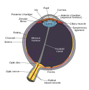
The eye is a complex biological device. The functioning of a camera is often compared with the workings of the eye, mostly since both focus light from external objects in the field of viewField of view
The field of view is the extent of the observable world that is seen at any given moment....
onto a light-sensitive medium. In the case of the camera, this medium is film or an electronic sensor; in the case of the eye, it is an array of visual receptors. With this simple geometrical similarity, based on the laws of optics, the eye functions as a transducer
Transducer
A transducer is a device that converts one type of energy to another. Energy types include electrical, mechanical, electromagnetic , chemical, acoustic or thermal energy. While the term transducer commonly implies the use of a sensor/detector, any device which converts energy can be considered a...
, as does a CCD camera
Charge-coupled device
A charge-coupled device is a device for the movement of electrical charge, usually from within the device to an area where the charge can be manipulated, for example conversion into a digital value. This is achieved by "shifting" the signals between stages within the device one at a time...
.
Light entering the eye is refracted as it passes through the cornea
Cornea
The cornea is the transparent front part of the eye that covers the iris, pupil, and anterior chamber. Together with the lens, the cornea refracts light, with the cornea accounting for approximately two-thirds of the eye's total optical power. In humans, the refractive power of the cornea is...
. It then passes through the pupil
Pupil
The pupil is a hole located in the center of the iris of the eye that allows light to enter the retina. It appears black because most of the light entering the pupil is absorbed by the tissues inside the eye. In humans the pupil is round, but other species, such as some cats, have slit pupils. In...
(controlled by the iris
Iris (anatomy)
The iris is a thin, circular structure in the eye, responsible for controlling the diameter and size of the pupils and thus the amount of light reaching the retina. "Eye color" is the color of the iris, which can be green, blue, or brown. In some cases it can be hazel , grey, violet, or even pink...
) and is further refracted by the lens. The cornea and lens act together as a compound lens to project an inverted image onto the retina.
Retina
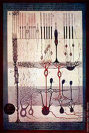
The retina consists of a large number of photoreceptor cells which contain particular protein
Protein
Proteins are biochemical compounds consisting of one or more polypeptides typically folded into a globular or fibrous form, facilitating a biological function. A polypeptide is a single linear polymer chain of amino acids bonded together by peptide bonds between the carboxyl and amino groups of...
molecule
Molecule
A molecule is an electrically neutral group of at least two atoms held together by covalent chemical bonds. Molecules are distinguished from ions by their electrical charge...
s called opsin
Opsin
Opsins are a group of light-sensitive 35–55 kDa membrane-bound G protein-coupled receptors of the retinylidene protein family found in photoreceptor cells of the retina. Five classical groups of opsins are involved in vision, mediating the conversion of a photon of light into an electrochemical...
s. In humans, two types of opsins are involved in conscious vision: rod opsins
Rod cell
Rod cells, or rods, are photoreceptor cells in the retina of the eye that can function in less intense light than can the other type of visual photoreceptor, cone cells. Named for their cylindrical shape, rods are concentrated at the outer edges of the retina and are used in peripheral vision. On...
and cone opsins
Cone cell
Cone cells, or cones, are photoreceptor cells in the retina of the eye that are responsible for color vision; they function best in relatively bright light, as opposed to rod cells that work better in dim light. If the retina is exposed to an intense visual stimulus, a negative afterimage will be...
. (A third type, melanopsin
Melanopsin
Melanopsin is a photopigment found in specialized photosensitive ganglion cells of the retina that are involved in the regulation of circadian rhythms, pupillary light reflex, and other non-visual responses to light. In structure, melanopsin is an opsin, a retinylidene protein variety of...
in some of the retinal ganglion cells (RGC), part of the body clock mechanism, is probably not involved in conscious vision, as these RGC do not project to the lateral geniculate nucleus
Lateral geniculate nucleus
The lateral geniculate nucleus is the primary relay center for visual information received from the retina of the eye. The LGN is found inside the thalamus of the brain....
(LGN) but to the pretectal olivary nucleus (PON).) An opsin absorbs a photon
Photon
In physics, a photon is an elementary particle, the quantum of the electromagnetic interaction and the basic unit of light and all other forms of electromagnetic radiation. It is also the force carrier for the electromagnetic force...
(a particle of light) and transmits a signal to the cell
Cell (biology)
The cell is the basic structural and functional unit of all known living organisms. It is the smallest unit of life that is classified as a living thing, and is often called the building block of life. The Alberts text discusses how the "cellular building blocks" move to shape developing embryos....
through a signal transduction pathway, resulting in hyperpolarization of the photoreceptor. (For more information, see Photoreceptor cell).
Rods and cones differ in function. Rods are found primarily in the periphery of the retina and are used to see at low levels of light. Cones are found primarily in the center (or fovea
Fovea
The fovea centralis, also generally known as the fovea , is a part of the eye, located in the center of the macula region of the retina....
) of the retina. There are three types of cones that differ in the wavelengths of light they absorb; they are usually called short or blue, middle or green, and long or red. Cones are used primarily to distinguish color
Color
Color or colour is the visual perceptual property corresponding in humans to the categories called red, green, blue and others. Color derives from the spectrum of light interacting in the eye with the spectral sensitivities of the light receptors...
and other features of the visual world at normal levels of light.
In the retina, the photoreceptors synapse directly onto bipolar cells, which in turn synapse onto ganglion cell
Ganglion cell
A retinal ganglion cell is a type of neuron located near the inner surface of the retina of the eye. It receives visual information from photoreceptors via two intermediate neuron types: bipolar cells and amacrine cells...
s of the outermost layer, which will then conduct action potentials to the brain
Brain
The brain is the center of the nervous system in all vertebrate and most invertebrate animals—only a few primitive invertebrates such as sponges, jellyfish, sea squirts and starfishes do not have one. It is located in the head, usually close to primary sensory apparatus such as vision, hearing,...
. A significant amount of visual processing arises from the patterns of communication between neuron
Neuron
A neuron is an electrically excitable cell that processes and transmits information by electrical and chemical signaling. Chemical signaling occurs via synapses, specialized connections with other cells. Neurons connect to each other to form networks. Neurons are the core components of the nervous...
s in the retina. About 130 million photoreceptors absorb light, yet roughly 1.2 million axons of ganglion cells transmit information from the retina to the brain. The processing in the retina includes the formation of center-surround receptive fields of bipolar and ganglion cells in the retina, as well as convergence and divergence from photoreceptor to bipolar cell. In addition, other neurons in the retina, particularly horizontal
Horizontal cell
Horizontal cells are the laterally interconnecting neurons in the outer plexiform layer of the retina of mammalian eyes. They help integrate and regulate the input from multiple photoreceptor cells...
and amacrine cell
Amacrine cell
Amacrine cells are interneurons in the retina. Amacrine cells are responsible for 70% of input to retinal ganglion cells. Bipolar cells, which are responsible for the other 30% of input to retinal ganglia, are regulated by amacrine cells.-Overview:...
s, transmit information laterally (from a neuron in one layer to an adjacent neuron in the same layer), resulting in more complex receptive fields that can be either indifferent to color and sensitive to motion
Motion (physics)
In physics, motion is a change in position of an object with respect to time. Change in action is the result of an unbalanced force. Motion is typically described in terms of velocity, acceleration, displacement and time . An object's velocity cannot change unless it is acted upon by a force, as...
or sensitive to color and indifferent to motion.
Mechanism of generating visual signals: The retina adapts to its change in light through the use of the rods. In the dark, the retinal has a bent shape called cis-retinal. When light is present, the retinal changes to a straight form called trans-retinal and breaks away from the opsin. This is called bleaching because the purified rhodopsin changes from violet to colorless in the light. In the dark, the rhodopsin absorbs no light therefore releasing glutamate cells which inhibit the bipolar cell. This inhibits the release of neurotransmitters to the ganglion cell. In the light, glutamate secretion ceases which no longer inhibits the bipolar cell from releasing neurotransmitters to the ganglion cell and therefore an image can be detected.
The final result of all this processing is five different populations of ganglion cells that send visual (image-forming and non-image-forming) information to the brain:
- M cells, with large center-surround receptive fields that are sensitive to depthDepth perceptionDepth perception is the visual ability to perceive the world in three dimensions and the distance of an object. Depth sensation is the ability to move accurately, or to respond consistently, based on the distances of objects in an environment....
, indifferent to color, and rapidly adapt to a stimulus; - P cells, with smaller center-surround receptive fields that are sensitive to color and shapeShapeThe shape of an object located in some space is a geometrical description of the part of that space occupied by the object, as determined by its external boundary – abstracting from location and orientation in space, size, and other properties such as colour, content, and material...
; - K cells, with very large center-only receptive fields that are sensitive to color and indifferent to shape or depth;
- another population that is intrinsically photosensitivePhotosensitive ganglion cellPhotosensitive ganglion cells, also called photosensitive Retinal Ganglion Cells , intrinsically photosensitive Retinal Ganglion Cells or melanopsin-containing ganglion cells, are a type of neuron in the retina of the mammalian eye.They were discovered in the early 1990sand are, unlike other...
; and - a final population that is used for eye movements.
A 2006 University of Pennsylvania
University of Pennsylvania
The University of Pennsylvania is a private, Ivy League university located in Philadelphia, Pennsylvania, United States. Penn is the fourth-oldest institution of higher education in the United States,Penn is the fourth-oldest using the founding dates claimed by each institution...
study calculated the approximate bandwidth
Bandwidth (computing)
In computer networking and computer science, bandwidth, network bandwidth, data bandwidth, or digital bandwidth is a measure of available or consumed data communication resources expressed in bits/second or multiples of it .Note that in textbooks on wireless communications, modem data transmission,...
of human retinas to be about 8960 kilobits per second, whereas guinea pig
Guinea pig
The guinea pig , also called the cavy, is a species of rodent belonging to the family Caviidae and the genus Cavia. Despite their common name, these animals are not in the pig family, nor are they from Guinea...
retinas transfer at about 875 kilobits.
In 2007 Zaidi and co-researchers on both sides of the Atlantic studying patients without rods and cones, discovered that the novel photoreceptive ganglion cell in humans also has a role in conscious and unconscious visual perception. The peak spectral sensitivity was 481 nm. This shows that there are two pathways for sight in the retina – one based on classic photoreceptors (rods and cones) and the other, newly discovered, based on photoreceptive ganglion cells which act as rudimentary visual brightness detectors.
Photochemistry
In the visual system, retinal, technically called retinene1 or "retinaldehyde", is a light-sensitive retineneRetinene
The Retinenes are chemical derivatives of the dietary supplement vitamin A formed through oxidation reactions....
molecule found in the rods and cones of the retina
Retina
The vertebrate retina is a light-sensitive tissue lining the inner surface of the eye. The optics of the eye create an image of the visual world on the retina, which serves much the same function as the film in a camera. Light striking the retina initiates a cascade of chemical and electrical...
. Retinal is the fundamental structure involved in the transduction of light
Light
Light or visible light is electromagnetic radiation that is visible to the human eye, and is responsible for the sense of sight. Visible light has wavelength in a range from about 380 nanometres to about 740 nm, with a frequency range of about 405 THz to 790 THz...
into visual signals, i.e. nerve impulses in the ocular system of the central nervous system
Central nervous system
The central nervous system is the part of the nervous system that integrates the information that it receives from, and coordinates the activity of, all parts of the bodies of bilaterian animals—that is, all multicellular animals except sponges and radially symmetric animals such as jellyfish...
. In the presence of light, the retinal molecule changes configuration and as a result a nerve impulse is generated.
Optic nerve
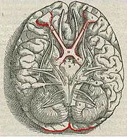
Optic nerve
The optic nerve, also called cranial nerve 2, transmits visual information from the retina to the brain. Derived from the embryonic retinal ganglion cell, a diverticulum located in the diencephalon, the optic nerve doesn't regenerate after transection.-Anatomy:The optic nerve is the second of...
. Different populations of ganglion cells in the retina send information to the brain through the optic nerve. About 90% of the axons in the optic nerve go to the lateral geniculate nucleus
Lateral geniculate nucleus
The lateral geniculate nucleus is the primary relay center for visual information received from the retina of the eye. The LGN is found inside the thalamus of the brain....
in the thalamus
Thalamus
The thalamus is a midline paired symmetrical structure within the brains of vertebrates, including humans. It is situated between the cerebral cortex and midbrain, both in terms of location and neurological connections...
. These axons originate from the M, P, and K ganglion cells in the retina, see above. This parallel processing is important for reconstructing the visual world; each type of information will go through a different route to perception
Perception
Perception is the process of attaining awareness or understanding of the environment by organizing and interpreting sensory information. All perception involves signals in the nervous system, which in turn result from physical stimulation of the sense organs...
. Another population sends information to the superior colliculus
Superior colliculus
The optic tectum or simply tectum is a paired structure that forms a major component of the vertebrate midbrain. In mammals this structure is more commonly called the superior colliculus , but, even in mammals, the adjective tectal is commonly used. The tectum is a layered structure, with a...
in the midbrain, which assists in controlling eye movements (saccades) as well as other motor responses.
A final population of photosensitive ganglion cell
Photosensitive ganglion cell
Photosensitive ganglion cells, also called photosensitive Retinal Ganglion Cells , intrinsically photosensitive Retinal Ganglion Cells or melanopsin-containing ganglion cells, are a type of neuron in the retina of the mammalian eye.They were discovered in the early 1990sand are, unlike other...
s, containing melanopsin
Melanopsin
Melanopsin is a photopigment found in specialized photosensitive ganglion cells of the retina that are involved in the regulation of circadian rhythms, pupillary light reflex, and other non-visual responses to light. In structure, melanopsin is an opsin, a retinylidene protein variety of...
, sends information via the retinohypothalamic tract
Retinohypothalamic tract
The retinohypothalamic tract is a photic input pathway involved in the circadian rhythms of mammals. The origin of the retinohypothalamic tract is the intrinsically photosensitive retinal ganglion cells , which contain the photopigment melanopsin...
(RHT) to the pretectum
Pretectum
The pretectum, also known as the pretectal area, is a region of neurons found between the thalamus and midbrain. It receives binocular sensory input from retinal ganglion cells of the eyes, and is the region responsible for maintaining the pupillary light reflex.-Outputs:The pretectum, after...
(pupillary reflex), to several structures involved in the control of circadian rhythms and sleep
Sleep
Sleep is a naturally recurring state characterized by reduced or absent consciousness, relatively suspended sensory activity, and inactivity of nearly all voluntary muscles. It is distinguished from quiet wakefulness by a decreased ability to react to stimuli, and is more easily reversible than...
such as the suprachiasmatic nucleus
Suprachiasmatic nucleus
The suprachiasmatic nucleus or nuclei, abbreviated SCN, is a tiny region on the brain's midline, situated directly above the optic chiasm. It is responsible for controlling circadian rhythms...
(SCN, the biological clock), and to the ventrolateral preoptic nucleus
Ventrolateral preoptic nucleus
The ventrolateral preoptic nucleus is a group of neurons in the hypothalamus. They are primarily active during Non-rapid eye movement sleep, and inhibit other neurons that are involved in wakefulness...
(VLPO, a region involved in sleep regulation). A recently discovered role for photoreceptive ganglion cells is that they mediate conscious and unconscious vision – acting as rudimentary visual brightness detectors as shown in rodless coneless eyes.
Optic chiasm
The optic nerves from both eyes meet and cross at the optic chiasm, at the base of the hypothalamusHypothalamus
The Hypothalamus is a portion of the brain that contains a number of small nuclei with a variety of functions...
of the brain. At this point the information coming from both eyes is combined and then splits according to the visual field
Visual field
The term visual field is sometimes used as a synonym to field of view, though they do not designate the same thing. The visual field is the "spatial array of visual sensations available to observation in introspectionist psychological experiments", while 'field of view' "refers to the physical...
. The corresponding halves of the field of view (right and left) are sent to the left and right halves of the brain, respectively, to be processed. That is, the right side of primary visual cortex deals with the left half of the field of view from both eyes, and similarly for the left brain. A small region in the center of the field of view is processed redundantly by both halves of the brain.
Optic tract
Information from the right visual field (now on the left side of the brain) travels in the left optic tract. Information from the left visual field travels in the right optic tract. Each optic tract terminates in the lateral geniculate nucleusLateral geniculate nucleus
The lateral geniculate nucleus is the primary relay center for visual information received from the retina of the eye. The LGN is found inside the thalamus of the brain....
(LGN) in the thalamus.
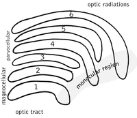
Lateral geniculate nucleus
The lateral geniculate nucleus (LGN) is a sensory relay nucleus in the thalamus of the brain. The LGN consists of six layers in humanHuman
Humans are the only living species in the Homo genus...
s and other primate
Primate
A primate is a mammal of the order Primates , which contains prosimians and simians. Primates arose from ancestors that lived in the trees of tropical forests; many primate characteristics represent adaptations to life in this challenging three-dimensional environment...
s starting from catarhinians, including cercopithecidae and apes. Layers 1, 4, and 6 correspond to information from the contralateral (crossed) fibers of the nasal visual field; layers 2, 3, and 5 correspond to information
Information
Information in its most restricted technical sense is a message or collection of messages that consists of an ordered sequence of symbols, or it is the meaning that can be interpreted from such a message or collection of messages. Information can be recorded or transmitted. It can be recorded as...
from the ipsilateral (uncrossed) fibers of the temporal visual field. Layer one (1) contains M cells which correspond to the M (magnocellular) cells of the optic nerve of the opposite eye and are concerned with depth or motion. Layers four and six (4 & 6) of the LGN also connect to the opposite eye, but to the P cells (color and edges) of the optic nerve. By contrast, layers two, three and five (2, 3, & 5) of the LGN connect to the M cells and P (parvocellular) cells of the optic nerve for the same side of the brain as its respective LGN. Spread out, the six layers of the LGN are the area of a credit card
Credit card
A credit card is a small plastic card issued to users as a system of payment. It allows its holder to buy goods and services based on the holder's promise to pay for these goods and services...
and about three times its thickness. The LGN is rolled up into two ellipsoids about the size and shape of two small birds' eggs. In between the six layers are smaller cells that receive information from the K cells (color) in the retina. The neurons of the LGN then relay the visual image to the primary visual cortex (V1) which is located at the back of the brain (caudal end) in the occipital lobe
Occipital lobe
The occipital lobe is the visual processing center of the mammalian brain containing most of the anatomical region of the visual cortex. The primary visual cortex is Brodmann area 17, commonly called V1...
in and close to the calcarine sulcus. The LGN is not just a simple relay station but it is also a center for processing; it receives reciprocal input from the cortical and subcortical layers and reciprocal innervation from the visual cortex.

Optic radiation
The optic radiations, one on each side of the brain, carry information from the thalamic lateral geniculate nucleusLateral geniculate nucleus
The lateral geniculate nucleus is the primary relay center for visual information received from the retina of the eye. The LGN is found inside the thalamus of the brain....
to layer 4 of the visual cortex
Visual cortex
The visual cortex of the brain is the part of the cerebral cortex responsible for processing visual information. It is located in the occipital lobe, in the back of the brain....
. The P layer neurons of the LGN relay to V1 layer 4C β. The M layer neurons relay to V1 layer 4C α. The K layer neurons in the LGN relay to large neurons called blobs in layers 2 and 3 of V1.
There is a direct correspondence from an angular position in the field of view
Field of view
The field of view is the extent of the observable world that is seen at any given moment....
of the eye, all the way through the optic tract to a nerve position in V1.
At this juncture in V1, the image path ceases to be straightforward; there is more cross-connection within the visual cortex.
Visual cortex
Cerebellum
The cerebellum is a region of the brain that plays an important role in motor control. It may also be involved in some cognitive functions such as attention and language, and in regulating fear and pleasure responses, but its movement-related functions are the most solidly established...
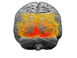
. The region that receives information directly from the LGN is called the primary visual cortex, (also called V1 and striate cortex). Visual information then flows through a cortical hierarchy. These areas include V2, V3, V4 and area V5/MT (the exact connectivity depends on the species of the animal). These secondary visual areas (collectively termed the extrastriate visual cortex) process a wide variety of visual primitives. Neurons in V1 and V2 respond selectively to bars of specific orientations, or combinations of bars. These are believed to support edge and corner detection. Similarly, basic information about color and motion is processed here.
Visual association cortex
As visual information passes forward through the visual hierarchy, the complexity of the neural representations increase. Whereas a V1 neuron may respond selectively to a line segment of a particular orientation in a particular retinotopic location, neurons in the lateral occipital complex respond selectively to complete object (e.g., a figure drawing), and neurons in visual association cortex may respond selectively to human faces, or to a particular object.Along with this increasing complexity of neural representation may come a level of specialization of processing into two distinct pathways: the dorsal stream and the ventral stream (the Two Streams hypothesis
Two Streams hypothesis
The two-streams hypothesis is a widely accepted, but still controversial, account of visual processing. As visual information exits the occipital lobe, it follows two main channels, or "streams". The ventral stream travels to the temporal lobe and is involved with object identification...
, first proposed by Ungerleider and Mishkin in 1982). The dorsal stream, commonly referred to as the "where" stream, is involved in spatial attention (covert and overt), and communicates with regions that control eye movements and hand movements. More recently, this area has been called the "how" stream to emphasize its role in guiding behaviors to spatial locations. The ventral stream, commonly referred as the "what" stream, is involved in the recognition, identification and categorization of visual stimuli.
However, there is still much debate about the degree of specialization within these two pathways, since they are in fact heavily interconnected.
See also
- EcholocationHuman echolocationHuman echolocation is the ability of humans to detect objects in their environment by sensing echoes from those objects. By actively creating sounds – for example, by tapping their canes, lightly stomping their foot or making clicking noises with their mouths – people trained to orientate with...
- Computer visionComputer visionComputer vision is a field that includes methods for acquiring, processing, analysing, and understanding images and, in general, high-dimensional data from the real world in order to produce numerical or symbolic information, e.g., in the forms of decisions...
- Helmholtz–Kohlrausch effectHelmholtz–Kohlrausch effectThe Helmholtz–Kohlrausch effect is an entoptic phenomenon wherein the intense saturation of spectral hue is perceived as part of the color's luminance...
- how color balanceColor balanceIn photography and image processing, color balance is the global adjustment of the intensities of the colors . An important goal of this adjustment is to render specific colors – particularly neutral colors – correctly; hence, the general method is sometimes called gray balance, neutral balance,...
affects vision - Memory-prediction frameworkMemory-prediction frameworkThe memory-prediction framework is a theory of brain function that was created by Jeff Hawkins and described in his 2004 book On Intelligence...
- Visual perceptionVisual perceptionVisual perception is the ability to interpret information and surroundings from the effects of visible light reaching the eye. The resulting perception is also known as eyesight, sight, or vision...
- Visual modularityVisual modularityIn cognitive neuroscience, visual modularity is an organizational concept concerning how vision works. The way in which the primate visual system operates is currently under intense scientific scrutiny...
Further reading
: the Google books link shows Alhazen's sketch of optic nerveOptic nerve
The optic nerve, also called cranial nerve 2, transmits visual information from the retina to the brain. Derived from the embryonic retinal ganglion cell, a diverticulum located in the diencephalon, the optic nerve doesn't regenerate after transection.-Anatomy:The optic nerve is the second of...
522 years before Vesalius
Vesalius
Andreas Vesalius was a Flemish anatomist, physician, and author of one of the most influential books on human anatomy, De humani corporis fabrica . Vesalius is often referred to as the founder of modern human anatomy. Vesalius is the Latinized form of Andries van Wesel...
' engraving....... (H.D. Steklis and J. Erwin, editors.) pp. 203–278... (References, pp. 180–198. Index, pp. 199–202. 202 pages.).
External links
- "Webvision: The Organization of the Retina and Visual System" – John Moran Eye Center at University of Utah
- VisionScience.com – An online resource for researchers in vision science.
- Journal of Vision – An online, open access journal of vision science.
- Hagfish research has found the “missing link” in the evolution of the eye. See: Nature Reviews Neuroscience.

