Tractography
Encyclopedia

Neuroscience
Neuroscience is the scientific study of the nervous system. Traditionally, neuroscience has been seen as a branch of biology. However, it is currently an interdisciplinary science that collaborates with other fields such as chemistry, computer science, engineering, linguistics, mathematics,...
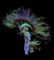
, tractography is a procedure to demonstrate the neural tracts.
It uses special techniques of magnetic resonance imaging
Magnetic resonance imaging
Magnetic resonance imaging , nuclear magnetic resonance imaging , or magnetic resonance tomography is a medical imaging technique used in radiology to visualize detailed internal structures...
(MRI), and computer-based image analysis
Image analysis
Image analysis is the extraction of meaningful information from images; mainly from digital images by means of digital image processing techniques...
.
The results are presented in two- and three-dimensional images.
In addition to the long tracts that connect the brain
Brain
The brain is the center of the nervous system in all vertebrate and most invertebrate animals—only a few primitive invertebrates such as sponges, jellyfish, sea squirts and starfishes do not have one. It is located in the head, usually close to primary sensory apparatus such as vision, hearing,...
to the rest of the body
Body
With regard to living things, a body is the physical body of an individual. "Body" often is used in connection with appearance, health issues and death...
, there is a complicated 3D network formed by short connections among different cortical
Cerebral cortex
The cerebral cortex is a sheet of neural tissue that is outermost to the cerebrum of the mammalian brain. It plays a key role in memory, attention, perceptual awareness, thought, language, and consciousness. It is constituted of up to six horizontal layers, each of which has a different...
and subcortical regions. The existence of these bundles has been revealed by histochemistry and biological
Biology
Biology is a natural science concerned with the study of life and living organisms, including their structure, function, growth, origin, evolution, distribution, and taxonomy. Biology is a vast subject containing many subdivisions, topics, and disciplines...
techniques on post-mortem specimens. Brain tracts are not identifiable by direct exam, CT
Computed tomography
X-ray computed tomography or Computer tomography , is a medical imaging method employing tomography created by computer processing...
, or MRI scans. This difficulty explains the paucity of their description in neuroanatomy
Neuroanatomy
Neuroanatomy is the study of the anatomy and organization of the nervous system. In contrast to animals with radial symmetry, whose nervous system consists of a distributed network of cells, animals with bilateral symmetry have segregated, defined nervous systems, and thus we can begin to speak of...
atlases and the poor understanding of their functions.
The MRI sequences used look at the symmetry of brain water diffusion. Bundles of fiber tracts make the water diffuse asymmetrically in a tensor
Tensor
Tensors are geometric objects that describe linear relations between vectors, scalars, and other tensors. Elementary examples include the dot product, the cross product, and linear maps. Vectors and scalars themselves are also tensors. A tensor can be represented as a multi-dimensional array of...
, the major axis parallel to the direction of the fibers. The asymmetry here is called anisotropy
Anisotropy
Anisotropy is the property of being directionally dependent, as opposed to isotropy, which implies identical properties in all directions. It can be defined as a difference, when measured along different axes, in a material's physical or mechanical properties An example of anisotropy is the light...
. There is a direct relationship between the number of fibers and the degree of anisotropy.

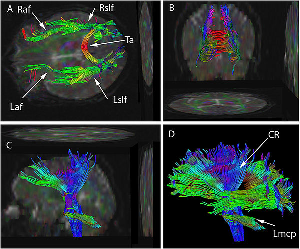
Figure legend:
Diffusion tensor imaging (DTI) data has been used to seed various tractographic assessments of this patient's brain. These are seen in superior (A), posterior (B), and lateral views (C&D). The seeds have been used to develop arcuate and superior longitudinal fasciculi in (A) and (B), for brainstem, and corona radiata in (C), and as combined data sets in (D). Some of the two dimensional projections of the tractographic result are also shown. The data set may be rotated continuously into various planes to better appreciate the structure. Color has been assigned based on the dominant direction of the fibers. There is asymmetry in the tractographic fiber volume between the right and left arcuate fasciculus (Raf & Laf) (smaller on the left) and between the right and left superior longitudinal fasciculus (Rslf & Lslf) (smaller on the right). Also seen are Tapetum (Ta), Left corona radiata (Lcr) and Left middle cerebellar peduncle (Lmcp).
MRI technique

Diffusion
Molecular diffusion, often called simply diffusion, is the thermal motion of all particles at temperatures above absolute zero. The rate of this movement is a function of temperature, viscosity of the fluid and the size of the particles...
of water
Water
Water is a chemical substance with the chemical formula H2O. A water molecule contains one oxygen and two hydrogen atoms connected by covalent bonds. Water is a liquid at ambient conditions, but it often co-exists on Earth with its solid state, ice, and gaseous state . Water also exists in a...
in the body, and can be used to reveal its 3D shape. Free diffusion occurs equally in all directions. This is termed "isotropic" diffusion. If the water diffuses in a medium with barriers, the diffusion will be uneven, which is termed "anisotropic" diffusion. In such a case, the relative mobility of the molecules from the origin has a shape different from a sphere
Sphere
A sphere is a perfectly round geometrical object in three-dimensional space, such as the shape of a round ball. Like a circle in two dimensions, a perfect sphere is completely symmetrical around its center, with all points on the surface lying the same distance r from the center point...
. This shape is often modeled as an ellipsoid, and the technique is then called Diffusion Tensor Imaging. Barriers can be many things--cell membranes, axons, myelin, etc; but in white matter the principal barrier is the myelin
Myelin
Myelin is a dielectric material that forms a layer, the myelin sheath, usually around only the axon of a neuron. It is essential for the proper functioning of the nervous system. Myelin is an outgrowth of a type of glial cell. The production of the myelin sheath is called myelination...
sheath of axons. Bundles of axons provide a barrier to perpendicular diffusion and a path for parallel diffusion along the orientation of the fibers.
Anisotropic diffusion is expected to be increased in areas of high mature axonal order. Conditions where the myelin or the structure of the axon are disrupted, such as trauma
Physical trauma
Trauma refers to "a body wound or shock produced by sudden physical injury, as from violence or accident." It can also be described as "a physical wound or injury, such as a fracture or blow." Major trauma can result in secondary complications such as circulatory shock, respiratory failure and death...
, tumors, and inflammation
Inflammation
Inflammation is part of the complex biological response of vascular tissues to harmful stimuli, such as pathogens, damaged cells, or irritants. Inflammation is a protective attempt by the organism to remove the injurious stimuli and to initiate the healing process...
reduce anisotropy, as the barriers are affected by destruction or disorganization.
Anisotropy is measured in several ways. One way is by a ratio called "fractional anisotropy
Fractional anisotropy
Fractional anisotropy is a scalar value between zero and one that describes the degree of anisotropy of a diffusion process. A value of zero means that diffusion is isotropic, i.e. it is unrestricted in all directions. A value of one means that diffusion occurs only along one axis and is fully...
" (FA). An anisotropy of "0" corresponds to a perfect sphere, whereas 1 is an ideal linear diffusion. Well-defined tracts have FA larger than 0.20. Few regions have FA larger than 0.90. The number gives information of how aspherical the diffusion is but says nothing of the direction.
Each anisotropy is linked to an orientation of the predominant axis (predominant direction of the diffusion). Post-processing programs are able to extract this directional information.
This additional information is difficult to represent on 2D grey-scaled images. To overcome this problem a color code is introduced . Basic colors can tell the observer how the fibers are oriented in a 3D-coordinate system: This is termed an "anisotropic map". The software could encode the colors in this way:
- Red indicates directions in the X axis: right to left or left to right.
- Green indicates directions in the Y axis: posterior to anterior or from anterior to posterior.
- Blue indicates directions in the Z axis: foot-to-head direction or vice versa
Notice that the technique is unable to discriminate the "positive" or "negative" direction in the same axis.

